Image-assisted therapy and intervention
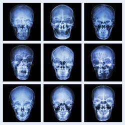
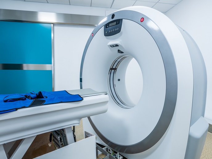
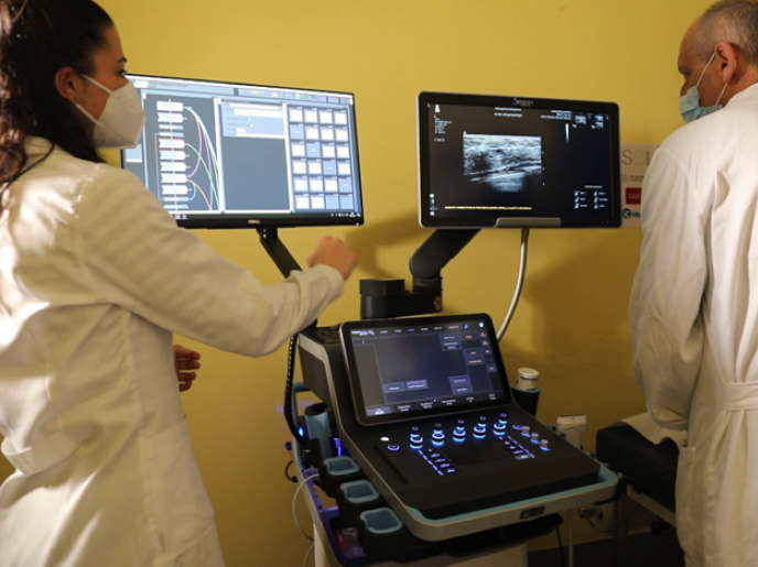
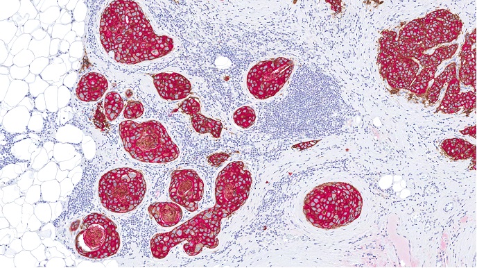
Recent advances in imaging provide unprecedented detail and resolution of body parts, thereby changing the way therapeutic and intervention procedures are executed. In addition, medical imaging could help predict and monitor recovery and prognosis after surgery as well as precisely provide the boundaries between diseased and healthy tissue. The latter often requires the combination of multimodal images for maximum resolution. The EU-funded TAHITI (Improving therapy and intervention through imaging) project was a multidisciplinary initiative with an overall aim to develop new techniques and methodologies to assist in the development of new therapeutic surgical procedures. The TAHITI consortium comprised three European institutions and Johns Hopkins University in the United States. Researchers generated tools that facilitate new image processing and image-guided procedures in cardiac therapy surgery planning. With respect to the assessment of myocardial infarction, they developed a new method for post-infarct scar segmentation and analysis of phase-sensitive inversion recovery images. They also proposed a novel protocol and analysis tools for magnetic resonance imaging (MRI) to determine area at risk, and a new method to obtain intuitive information for electrophysiology procedures. Project activities also included methodology development for liver surgery planning. Cystoscopy tools were tested in the fields of liver therapy and surgery as well as in endoscopy procedures. The combination of computed tomography and MRI data sets facilitated the diagnosis of metastases and segmentation of healthy parenchyma, thereby aiding surgical interventions accordingly. Additionally, multimodal combinations of 3D preoperative data with real-time monitoring during the procedure greatly helped soft-tissue surgery and abdominal interventions. Furthermore, imaging helped to delineate the in vivo effects of new drugs in causing chemotherapy-induced peripheral neuropathy. Taken together, the deliverables of the TAHITI project are expected to improve the healthcare of European citizens by providing new and safer means of therapy and surgery.


