Colour vision mechanisms illuminated
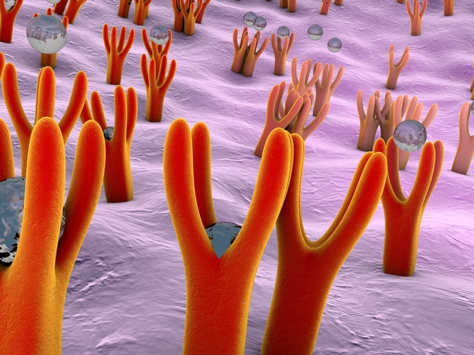
The retina of vertebrates contains the cells responsible for colour, rods and, more prominently, cones with blue, red and green pigment. Upon photoactivation, these pigments set off the visual signalling phototransduction cascade whereby light is converted into electrical signals in the cells of the retina to form the functional visual pigment rhodopsin. Mutations in the genes for cone pigments cause cone degeneration. The CONESYSTEM (Vision in color: Molecular mechanism of the color visual system) project has looked at the molecular basis behind the deterioration of colour vision due to cone cell dystrophies. The researchers expressed the individual proteins and characterised them with spectroscopic techniques. Due to its importance in the research as little was known about cone transducin, they determined the activity of this purified protein with the GTPγS35 binding assay. They also used surface plasmon resonance to investigate the kinetics of its molecular binding. However, the scientists still have work to do to fully understand how cone transducin binds with its receptor. CONESYSTEM researchers have successfully characterised all pigments involved in colour vision, each of which is responsible for a specific wavelength range. This is significant as the structure-function relationship in the least-known human photopigment, transducin, has been determined.


