New techniques to study repair of broken DNA
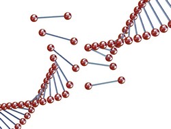
Elucidating the DNA repair process is critical for developing effective therapies. Scientists working on the SM-DNA-REPAIR (New single-molecule techniques and their application in the study of DNA break repair) project developed biophysical instruments and techniques to visualise the real-time dynamics of DNA repair at the single molecule level. Researchers focussed on the AddAB helicase-nuclease and the structural maintenance of chromosomes (SMC) complex from Bacillus subtilis. These complexes are implicated in DNA repair and help to maintain genome integrity. Any disruptions in their function could result in life-threatening illnesses. Several significant milestones were accomplished during the course of the project. Scientists built an optical and magnetic tweezers setup using an optical microscope, a CCD camera and magnetic beads. This system was used to determine the dynamics of AddAB translocation and the crucial role of regulatory sequences during double-stranded DNA break resection. In particular, studies provided novel insight into the mechanisms of crossover hotspot instigator (Chi) recognition, which are specific recombination hotspot sequences. Results were published in Molecular Cell in 2011 and PNAS in 2013. SM-DNA-REPAIR members also studied DNA-protein interactions at the single particle level after developing an optical tweezers setup involving video microscopy and lasers. This enabled researchers to determine the forces involved in single biotin-strepavidin interactions as well as to measure the force-extension curves of double stranded DNA and RNA. Results were published in JACS in 2013. Researchers successfully modified a commercial atomic force microscope (AFM) for imaging DNA in liquid buffer and air at an impressive rate of one image per second. Using this system, researchers were able to obtain deeper understanding into the AddAB resection reaction as well as the stoichiometry of protein complexes such as SMC. Project members' expertise in AFM imaging proved invaluable in several research collaborations with results being published in many high impact peer-reviewed journals. Topics included dynamics of double-stranded RNA, nanostructure characterisation and peptide binding to DNA. The SM-DNA-REPAIR tools and methodologies have applications that go way beyond the study of mechanisms involved in DNA repair. These tools could prove invaluable in tissue engineering, protein engineering as well as drug and biomaterials development. Moreover, these can also be adapted to research geared towards enhancing food security through better crop breeding and animal husbandry.