Virtual 3D organs help train surgeons
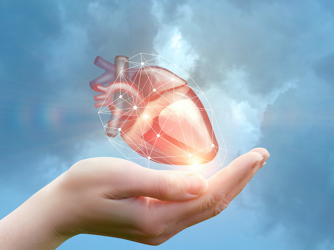
Millions of people require early diagnosis and urgent medical attention due to dangerous diseases of the heart and other organs. Doctors and surgeons working in this area must make rapid diagnosis and often perform emergency surgery. To improve current interventions, surgeons must learn through the use of medical corpses, which are difficult to access. “The medical field has extensive use of different scans such as X-ray, CT(opens in new window), MRI(opens in new window) and ultrasound. These techniques are very useful for providing information on internal organs. However, there is a gap in obtaining detailed, high-resolution organ information,” explains website in Italian(opens in new window) (Riccardo Roggeri), director and chief scientific officer at MTM(opens in new window). The EU-funded ROG project has created a game-changing platform that can create 3D virtual models of organs, using state-of-the-art technologies including artificial intelligence, and virtual and augmented reality. “ROG will lead to a significant decrease in surgical errors resulting from the incorrect evaluation of diagnostic images. The average duration of cardiovascular surgery will be reduced by 20 %,” says Roggeri, ROG project coordinator.
Intelligent algorithms at the heart of the software
The platform is based on a group of algorithms able to capture 2D images and transform them into 3D shapes. The team has already tested and validated the system on heart images, to generate hyperrealistic models with resolutions over 10 times higher than those achieved by current medical imaging today. The innovative software can take images of organs and tissues from medical scanning devices and let users analyse the resulting 3D models at different anatomical levels and regions. The programme can even recognise anomalies itself, identifying mutations and aberrations in morphology. “ROG analyses the density of points of 3D images and compares it against a database of tissues and organic structures. In this way it is easy to identify some cardiac and cardiovascular problems, such as calcifications(opens in new window) and occlusions(opens in new window),” adds Roggeri. The platform lets users build up extensive libraries of both 2D images and 3D models; a vast database on which new surgeons can train and existing surgeons can improve. It also offers the possibility of greater awareness to patients, who will be able to see realistic models of their own heart and cardiovascular system. “In this way the patient will be more involved in the decision-making phase and more informed about their health,” notes Roggeri.
Modelling for the present and future
“For now, the 3D organ models have been conceived and developed to be used by surgeons in the preoperative phase or by trainee surgeons to learn intervention techniques,” says Roggeri. In the near future, through a phase II continuation of the programme, the team hope to use AI and blockchain technologies to advance the ROG system further. “It will be possible to create tailor-made organs in the 3D image database, which replace the patient’s diseased organs,” Roggeri explains. To get to this stage, the ROG team will require a huge number of images to improve the statistical and medical accuracy of the system. “For this reason, doctors and surgeons from all over Europe are invited to participate with their hospitals in the testing and development programme of the platform,” concludes Roggeri.


