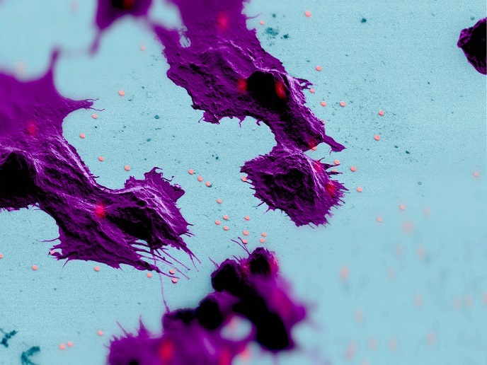
Live cell microscopy at set oxygen levels
When breathing, atmospheric oxygen is inhaled, which is then transported by the blood to the body to facilitate energy production. The normal oxygen levels in tissues range between 4-9 %, much lower than the atmospheric levels (21 %). Oxygen levels above (hyperoxia) or below (hypoxia) the normal threshold may be detrimental to cells and tissues. This is exemplified by ischaemia that may lead to heart attack or the acidification of muscles during exercise. Large solid cancers are also typically hypoxic due to poor vascularisation, strongly impeding the outcome of treatment.
A novel microscopy chamber for adjusting oxygen levels
Cell-based assays are routinely used in drug discovery processes, which are central to our society’s growing medical needs. The majority of these studies are carried out using live-cell microscopy methods with powerful fluorescent reporter dyes that are capable of visualising individual signals in living cells in real time and with high resolution. However, such studies are carried out at ambient oxygen conditions, which are considered hyperoxic for cells. Undertaken with the support of the Marie Skłodowska-Curie Actions(opens in new window) (MSCA) programme, the MICROX project proposed to develop an airtight microscopy chamber that would allow researchers to work at arbitrarily-set oxygen levels. “Our goal was to recapitulate the in vivo conditions of cells and tissues and make cell assays more relevant and accurate,” explains project coordinator Kees Jalink(opens in new window). It has proven quite difficult to achieve true hypoxic conditions on a microscope while at the same time assuring good access to the cells so as to be able to add hormones and drugs to study the signalling mechanisms. The proposed microscopy chamber was successfully developed and employed for routine experimentation at different oxygen levels. Using this setup, gas (oxygen, nitrogen, carbon dioxide) conditions can be changed within minutes, achieving absolute hypoxia with reliable oxygen levels below 0.5 %. The MICROX setup clearly outperforms available commercial solutions.
Cell signalling at various oxygen levels
Researchers also validated the functionality of available fluorescent sensors at hypoxia and investigated the impact of different oxygen levels on key cell signalling events. The work focused on adenosine 3’,5’-cyclic monophosphate(opens in new window) cAMP, a nucleotide that acts as a key second messenger in numerous signal transduction pathways converting extracellular signals received by cell surface receptors to intracellular signals. cAMP has a rapid life cycle and regulates various cellular functions, including cell growth and differentiation, gene transcription and protein expression. According to the MSCA research fellow Olga Kukk: “Although long-term hypoxia activates the hypoxia-inducible factor-1 and modulates gene transcription, our hypothesis was that the immediate (seconds to a few hours) effect of hypoxia on cell signalling would be transcription-independent.” Intriguingly, researchers were unable to verify this hypothesis in various fast signalling systems that they investigated, observing no apparent alterations of signals.
The promise of MICROX microscopy in drug discovery
The MICROX microscopy setup offers the capacity to fully adjust to atmospheric conditions, an important step in the establishment of physiological cell assays for screening biologically active compounds. Moreover, it is expected to contribute to the research investigation of dynamic events in living cells, which require exquisite temporal and spatial resolution.
Keywords
MICROX, oxygen levels, hypoxia, microscopy chamber, cAMP, hyperoxia, drug screening


