A microscope for early cancer detection
Long before tumour manifestation, tissue epithelial cells and surrounding stroma undergo molecular changes and alterations in tissue microarchitecture. These precancerous lesions or dysplasia imply that the tissue may be prone to develop cancer, but they cannot be easily detected by current imaging modalities. However, detection of such alterations has the power to improve cancer treatment prospects and patient survival.
Combining different modalities in one microscope
To address this diagnostic limitation, the EU-funded SENSITIVE(opens in new window) project developed a hybrid imaging modality that combines scattering for structural information and Raman spectroscopy(opens in new window) to quantitatively and qualitatively determine the molecular composition of a sample. These modalities were combined in a single microscope to facilitate ex vivo visualisation and discrimination between healthy and tumour tissue from excised thick tissue samples. Among the advantages of the generated imaging modalities is that they are label-free and hardly require any tissue processing, nor exogenous contrast agents like conventional histological evaluation by pathologists. Moreover, they can detect structural and molecular tissue changes which are not necessarily visible through traditional histopathological analysis. “Our methodology allows the identification of cancerous tissue or even dysplasia within tissue biopsies fresh from surgery,” explains project coordinator Apostolos Klinakis.
An endoscope for human use
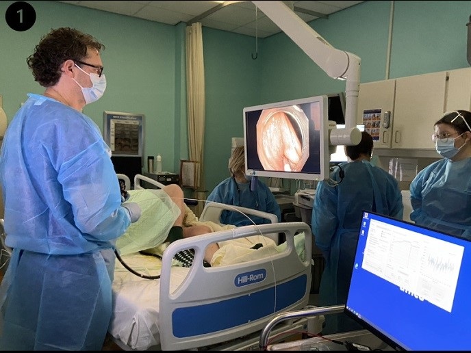
Despite challenges such as the COVID-19 pandemic, the consortium successfully designed and constructed a multimodal microscope and a clinical endoscope with Raman and scattering imaging modules. The SENSITIVE technology has been tested ex vivo on biopsies from oesophageal cancer and small intestine carcinomas from mouse models. Each modality alone, but more importantly combined, can discriminate between healthy tissue, tumour-adjacent non-diseased tissue and the tumour itself. The endoscope with Raman and scattering modalities has received institutional board approval for human studies. An ongoing clinical trial in Barrett oesophagus and Lynch syndrome patients has recruited the first 30 patients and shows promising results(opens in new window) along the same lines.
Diagnosis of field cancerisation
The SENSITIVE consortium also investigated the potential of applying their system for visualising field cancerisation(opens in new window) in whole tissue samples, ex vivo and in vivo, for detecting the presence of transformed cells adjacent to the primary tumour. Field cancerisation is characterised by the occurrence of genetic and epigenetic alterations in tissues that appear histologically normal and is believed to be a mediator of disease progression and relapse. Fuelled by the promising results, the anticipated next steps will be to complete the ongoing feasibility and safety clinical trial and hopefully find funds for a subsequent phase II clinical study, where the discrimination power of the methodology to detect field cancerisation will be assessed. The future of cancer medicine is geared towards prevention and early diagnosis. It will combine a plethora of genetic, lifestyle and molecular data to build individual patient profiles and calculate the risk of cancer development. The SENSITIVE system has the power to help stratify individuals into low- and high-risk groups so that they are regularly monitored for cancer detection at an early stage when cancer is still curable.