Tracing brain cancer’s ‘cell of origin’ points to pre-birth
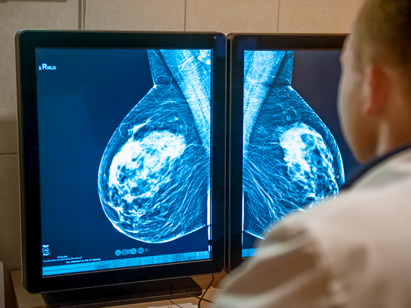
Compared to other cancers, brain tumours remain stubbornly difficult to treat. The blood-brain barrier prevents most therapeutics from reaching the brain, and as an immune-privileged organ, it is particularly difficult to leverage the immune system’s own defences in this area. In addition, the brain is vulnerable to long-term damage and life-threatening complications, requiring studies and treatments to be undertaken with caution. “Brain tumours are also extremely diverse, with probably around 200 different types, making each very rare,” says Stefan Pfister, project coordinator of BRAIN-MATCH(opens in new window), a project funded by the European Research Council(opens in new window). “Crucially, the fact that the cell of origin is also often unknown further impedes the development of specific treatments.” To help identify the cell of origin that gives rise to brain tumours, BRAIN-MATCH developed atlases to compare normal brain development in embryos and children with the molecular profiles of various brain tumour types in children, taken from data sets containing over 100 such tumours. “This told us about the timing and mechanisms by which the tumour hijacks normal processes, preventing it from being recognised by the body as ‘foreign’ and so a threat,” explains Pfister, from the German Cancer Research Center(opens in new window), the project’s host institution. Pfister is also affiliated with the Hopp Children’s Cancer Center Heidelberg(opens in new window) and Heidelberg University Hospital(opens in new window) in Germany.
Tracing the origin story
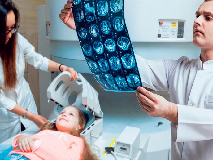
BRAIN-MATCH was inspired by the long-standing – but still unproven – hypothesis that childhood brain tumours are embryonal in origin, and may even be initiated in early pregnancy. Since the cellular origin of most brain tumours remains unknown, they are hard to model and to develop targeted treatments for, as these treatments must distinguish between the embryonal properties of tumour cells and normal differentiated brain tissues. The BRAIN-MATCH team used single-cell transcriptome(opens in new window) and ATAC sequencing(opens in new window) to analyse frozen brain tissue samples, starting at the embryonic stages. This resulted in large atlases of normal and cancerous brain development, specifically the cerebellum and brainstem, at single-cell resolution and characterising several hundred thousand cells. This was complemented by spatial transcriptome analyses, a molecular profiling method which can reconstruct tissues at the cellular level. “We were surprised how little is known about some cell types in the normal developing human brain, especially in the cerebellum and brainstem, where the majority of childhood brain tumours arise,” says Pfister. Analysis of both the cellular composition and differentiation of normal and tumour tissues highlighted commonalities and differences, ultimately pinpointing the cellular origin of various tumour types. “A key finding was that many tumours show extensive differentiation, from very primitive progenitor states all the way to differentiated cells, a progression very similar to normal cells,” notes Pfister. “This suggests cancerous cells hijack properties of normal cells during the embryonal period, rather than after birth.” He adds that the findings provide attractive targets for tissue-specific and time-bound treatments, minimising side effects. These properties could also help reliably identify tumour cells or nucleic acids in the cerebrospinal fluid or blood, enabling clinicians to make a diagnosis without operations or biopsies. It would also offer an effective means to monitor the response of patients to treatment.
A rich therapeutics resource
There is currently little financial incentive for industry to develop specific therapeutics for rare diseases such as childhood brain tumours. In addition, most drugs that could potentially be repurposed for these cancers are designed specifically not to penetrate the brain. “While therapeutic developments will probably need novel financing arrangements, our data offers a rich resource to help identify and prioritise therapeutic targets for these rare diseases. Our data set of normal human brain development is also relevant for research fields beyond cancer, such as cognitive health, brain injury and neurodegeneration,” concludes Pfister. After the relevant publications, the project’s brain atlases will be made available to researchers, with another brainstem atlas currently under development. Meanwhile, a paper on mouse atlases has already been published in ‘Science’(opens in new window), with another on the human cerebellum pending. Some of the results have also been recently published in ‘Neuro-Oncology’(opens in new window). The team is now focused on functionally validating the tissue specificity of the targets identified and their role in killing tumour cells. Various tumour types will also be modelled to understand their mechanisms of resistance during their evolution.