



Drawing 3D models of bone marrow with silk ink

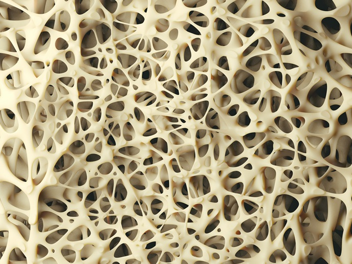
The human body possesses a remarkable ability to respond to injuries by producing platelets that facilitate blood clotting. This intricate process is led by giant cells called megakaryocytes that reside in the marrow of our bones. However, conditions such as leukaemia, viral infections and blood disease can interrupt this process, leaving the patient vulnerable to injury. Researchers with the SILKink(opens in new window) project, which was funded by the European Innovation Council(opens in new window), took on this challenge by developing an innovative bioink printing machine using silk as a base material. The system can 3D-print bone marrow models that can then be seeded with living cells to study bone disease and even to manufacture platelets ex vivo. This innovative advance could greatly enhance Europe’s medical research and lead to the production of tailored drugs and personalised medicine, a key aim of the EU4Health strategy(opens in new window). “The goal of the project was using silk ink to understand if we can use this system to study drugs,” says SILKink project coordinator Alessandra Balduini, from the Department of Molecular Medicine at the University of Pavia(opens in new window) in Italy.
Improved bone marrow models
Balduini and her team have developed bone marrow models for years, improving their mechanisms and sharpening clinical aspects. Any bioink used to create bone marrow models needs to provide sufficient structure to allow differentiation of megakaryocytes, the large cells in bone marrow that produce platelets. While existing models have functioned well, they lack the softness necessary to closely mimic bone marrow tissue. If bone marrow structure is not well represented, cells introduced to it can become stressed and damaged. This results in changes to their DNA that masks the effect of drugs being tested in the model, making it harder to identify good treatments. For this reason, the researchers turned to silk. A natural fibre harvested from the cocoons of domesticated silkworms, silk fibre is both flexible and incredibly strong, and is also an effective means of drug delivery. “Since we are mimicking the bone marrow, the cells are in a physiological-like state, so they have less DNA damage, they have less stress, less inflammation. So you can study the impact of a drug in a real system,” explains Balduini.
Commercialising the healing properties of silk
Another major outcome of the project was initiating a start-up, Silk4B(opens in new window), to commercialise the team’s advanced 3D bone marrow models and related silk-based products. “We give the possibility to researchers to use bone marrow models they can really trust for their experiments,” notes Balduini. The start-up is selling the products to researchers, pharmaceutical companies and academics worldwide.
Further development of the silk bioink
Balduini and her team continue to work on the ink, and have for example developed a way for it to change colour to give an easier read-out of any changes to the system. They are also working on a silk bone marrow platform to mass-produce platelets for transfusion. They are now studying different applications for the models, including culturing patient cells and seeing how they respond to drugs. Eventually, the hope is this silk-based system will be used in clinical settings, though this is still a few years off.