Unlocking brain tumour secrets to transform cancer treatment

Standard magnetic resonance imaging (MRI) scans show the size and location of brain tumours but reveal little about the biological properties that determine whether treatment will work. With support from the Marie Skłodowska-Curie Actions(opens in new window) (MSCA) programme, researcher Giacomo Annio used a sophisticated imaging technique called magnetic resonance elastography (MRE) to understand glioblastomas, the most aggressive brain tumours. “Elastography measures how stiff or soft [tumour] tissue is – properties that provide clues to tumour behaviour and how it resists therapy and infiltrates surrounding tissue,” he explains. Annio used glioblastoma data from Oslo University Hospital(opens in new window) (OUH), Norway, after conducting preclinical research at Stanford University, United States.
Device the size of a mobile phone
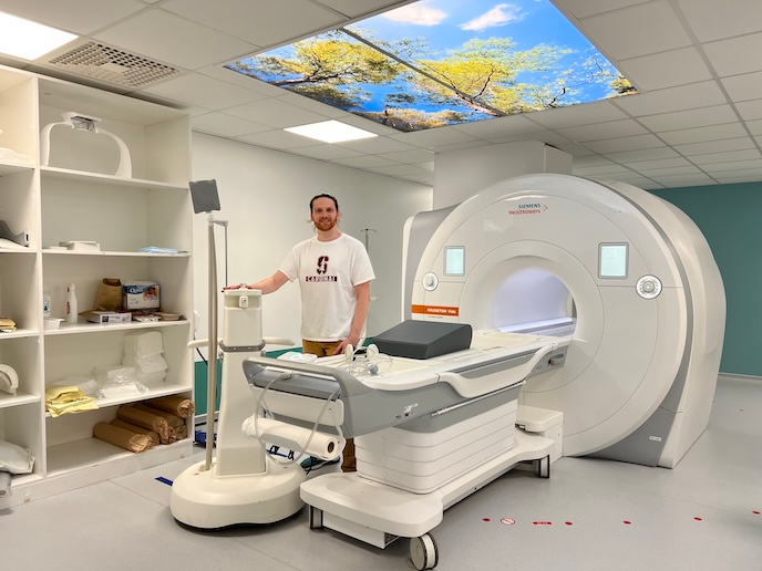
The vibrating MRE device, the size of a mobile phone, is placed against the patient’s head, sending waves into the brain. A standard MRI scanner visualises these waves as they travel through tissue. “It allows clinicians to look at the tumour in a new way: looking at their physical properties, providing a new perspective to understand tumour biology,” Annio says. The MRE device is currently being developed by American company Quality Electrodynamics and commercialised on MRI scanners by Siemens in Germany. It is beginning to appear in hospitals. But Annio remarks: “Adapting it for brain tumours is a particular challenge.” The EU-funded GLIOBID project builds on previous EU-funded projects: FORCE which developed MRE; and ImPRESS, applying MRE to brain tumours. Annio was part of both these teams.
Laboratory trials in the United States
Annio spent the first two years of his MSCA fellowship at Stanford’s Department of Radiology, Division of Molecular Imaging Program(opens in new window). He built an MRE device in the lab using 3D printing and customised electronics, the first of its kind at Stanford. It was installed on an MRI scanner to study brain tumours in mice. “At Stanford, we patented a theranostic agent – a molecule that combines therapy and diagnosis and can be visualised using MRI,” Annio notes. “This molecule releases a component that changes the mechanics of the tumour, opening up the blood vessels specifically at the tumour site.” He goes on to add that this would allow more treatment to flow inside the tumour. “Using elastography, we could see how the mechanical properties of the tumour impact on the delivery of therapy. No one had done this before.” Lesions shrank in mice treated with this additional therapy. “We increased survival,” according to Annio. This approach would enable a therapeutic response based on a tumour’s biomechanical fingerprint, sparing patients from ineffective treatments and advancing personalised treatment, he adds.
Shedding light on glioblastomas
With data from ongoing clinical trials at OUH involving 200 glioblastoma patients, MRE revealed more about these aggressive tumours. “While most cancers are stiffer than surrounding tissue, brain glioblastomas are softer,” Annio explains. But tumour viscosity, which he describes as ‘squeezability’, may matter more than stiffness in predicting tumour aggressiveness. MRE also sheds light on blood vessels inside the tumour and pressure that builds up inside the brain. As the tumour grows, tumour cells multiply, new scaffolding is built and new vessels form to deliver ‘fuel’ to the tumour, Annio notes. “In this crowded space, tumour blood vessels are squeezed, and they cannot properly deliver blood, oxygen, nutrients and, importantly, treatment.” Project coordinator Kyrre Eeg Emblem, head of the Department of Physics and Computational Radiology at OUH, says the project enabled research to be expanded to other types of brain tumour such as meningioma, a more common brain tumour. “Here, the assessment of physical forces allows the neurosurgeon to plan surgery by reviewing whether the tumour will be easy or difficult to remove,” he adds.