Detecting genetic abnormalities in real time using fluorescence
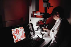
Our genetic make up, or DNA, consists of nucleic acids, coupled together with partner nucleic acids to form two chains, which are bonded together in the famous double helix structure. Genetic problems arise when these coupling reactions do not take place as they should. Present diagnostic systems used to detect any gene deformities, focus on coupling such nucleic acids with a probe nucleic acid, to create a hybrid. The probe contains some form of indicator or label, often a dye, which will allow the hybrid to be monitored and any abnormalities to be detected. Unfortunately this type of hybridization is limited with regard to sensitivity, specificity, complexity, hybridization conditions and the need for prior probe labelling. A new method of detecting abnormalities involves monitoring these hybrid reactions in real time. DNA molecules are attached to a gold-coated glass chip, which is then mounted onto a surface plasmon microscope and coupled to a flow through cell. The complementary acids, which are labelled with specific dye, are then passed through this flow through cell. The system is excited to a higher energy, and if the acids couple successfully, the extra energy is release. This fluorescene energy can be monitored on-line with a suitable imaging optical and very sensitive CCD camera. This system allows the parallel monitoring of simultaneous binding events. The images are transferred to a computer and image analysis software routines allow the coupling reactions to be quantified, their rates determined, and a single mismatch can be easily detected. This novel system has been proved in principle. Industrial partners are now being sought to assess applicability of the on-line chip read-out scheme.