Optimising radiation protection at low doses
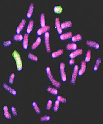
Nowadays, information on cancer risk after ionising radiation is more widely known in comparison to any other cause. This is mainly due to the fact that scientists have for many decades, extensively studied tumour development in people exposed to high and low doses. However, since cancer is a very complex disease to which multiple factors contribute, the associated risk at very low doses is still very difficult to estimate. Urged by this, the MAGELLANS project focused on studying the mechanisms and genetics of tumorigenesis afterradiation exposure. Within this context, researchers exploited mouse models of leukaemia, lymphoma and skin neoplasia and the useful data generated may provide the basis for determining human radiation cancer risk at low doses. Furthermore, these data coupled with knowledge of human genetics may help us to understand distribution of cancer risk within the population and whether individuals may be genetically susceptible. Both published (27 scientific papers) and unpublished data constitute supportive evidence that a gene loss mechanism plays a vital role in the early effects of radiation on multi-stage tumourigenesis in the skin and haemopoietic system. With other data this implies that even the lowest dose of ionising radiation, coming from natural or man-made sources, has increased the potential of developing malignancies. In addition, a few candidate variant genes such as Pthlh and Scaa2 for skin or Prkdc in the breast and intestine were identified as having roles in genetic susceptibility. The derived data may be further exploited to analyse the multiple ways by which various genetic risk determinants interact in different tumour types. Therefore, the project results may provide valuable input for cancer studies in terms of tumour development and early diagnosis. They may also contribute to the improvement of radiation protection standards on the health aspects of natural, industrial and medical uses of ionising radiation. The image shows Molecular staining (in green) of the chromosome number 2 copies of a cell from a radiation-induced acute myeloid leukaemia (AML). One copy is reduced in size by a radiation-associated deletion characteristic of AML.