Brain tissue characteristics diagnose prion diseases
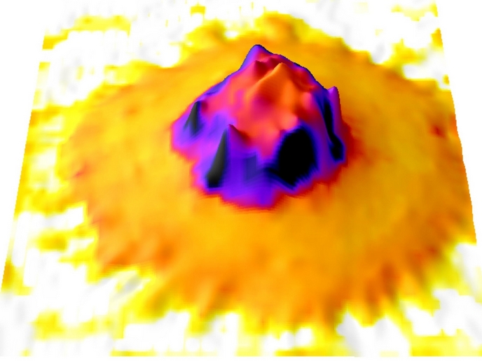
The appearance of the variant form of Creutzfeld-Jakob disease (CJD) fuelled research into possible therapies. Essential for this is a comprehensive knowledge of the mechanisms and stages involved during disease progression. Partners in the EU funded PRIONMRDIAGNOSTICS sought to find non-invasive magnetic resonance (MR) biomarkers for diagnostic criteria of human prion diseases. Determination of tissue characteristics using MR is used in the diagnosis of many diseases including those of the central nervous system. Ideal candidate pathologies are effects of strokes, many psychiatric disorders and the various forms of transmissible spongiform encephalopathies (TSEs). For this study, data was collected from patients and animal models with various forms of TSE. Specifically, individuals with sporadic or variant CJD, inherited forms of TSE and non-prion forms of dementia as well as two animal models were investigated using different modes of MR methodology. The project team at the University of Bologna focused on identifying new MR biomarkers that demonstrated the required specificity and sensitivity. Quantitative data from MR imaging (MRI) and MR spectroscopy (MRS) was collated and analysed statistically. Regional differences in brain tissue characteristics at various stages in the pathologies could then be determined. The team found that diagnostic protocols could be improved by the inclusion of proton MRS (1H-MRS) applied to the thalamus. Diffusion-weighted magnetic resonance imaging (DWI) was also recommended as a promising tool for understanding changes in brain tissue in cases of TSE. The findings of this research were disseminated through scientific journals. Direct application of MR indicators to patient care and further study through national CJD surveillance units was also planned through their inclusion in diagnostic procedures. Improved disease indicators mean that development and testing of novel therapies could also be facilitated.