Advanced imaging techniques for detecting pathology
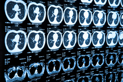
In vivo imaging techniques are widely used in the diagnosis of abnormalities in both structure and function. These techniques enable physicians to see two-dimensional (2D) slices or 3D reconstructions of a patient’s brain including indicators of its function over time in different tasks. The ‘Detection of brain abnormality’ (DEBRA) project has been designed to develop computer-assisted lesion segmentation techniques for enhanced CVD detection using MRI and with widespread application to neuroimaging of other pathologies. Specifically, the investigators are applying pattern classification techniques in combination with statistical modelling to enhance detection of subtle differences both in disease tissue with respect to healthy tissue and in disease tissue over time. To date, the researchers have collected clinical data, created simulated data, carried out pre-processing of imaging data and developed and implemented algorithms for abnormality segmentation. The algorithms provide enhanced sensitivity for both real and simulated datasets when compared to conventional methods. The specificity currently lacking is to be expected given the elimination of manual selection and a priori knowledge. Nevertheless, the algorithms to date could be used for expediting the screening process or in combination with a supervised scheme for increased accuracy. In addition, continuing project work should improve the specificity of the algorithms in abnormality detection and expand its general applicability to detecting pathologies other than those associated with CVD.