The devil is in the details
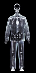
X-rays were first discovered and documented by the German scientist Wilhelm Roentgen. He observed the detailed images of the bones in his hands and arms after submitting them to streams of X-rays. Different materials absorb X-rays in different amounts. The structures that absorb the radiation to a greater degree produce darker images on X-ray film, sort of like shadows, indicating that fewer X-rays are detected by the film. Although the technology has been around for a long time, techniques have been extended to produce ever higher resolution images. One area of focus is the use of scintillators, or materials that emit visible light (become luminescent) after absorbing radiation (in this case, x-radiation), and scintillator detectors that essentially magnify the signal. Researchers designed the ‘Novel ceramic thin film based scintillator for high resolution X-ray imaging’ (Scintax) project to develop novel scintillating films and subsequently new X-ray detectors for scientific and industrial application. The researchers focused on epitaxially grown, doped Lu2SiO5 (LSO, lutetium orthosilicate) scintillators particularly well suited to indirect X-ray detection via a visible light detector. Experiments resulted in publication of standard characterisation methods for digital detector arrays as well as a patent application. The Scintax project systems are well suited to non-destructive inspection, providing enhanced safety due to higher spatial resolution. In addition, enhanced resolution even at higher energies and reduced exposure times opens the door for more widespread applications in medicine and biology. Commercialisation has the potential to enhance the safety of all sorts of products subjected to non-destructive testing. Equally important, the new technology just might make early detection of disease states easier and thus recovery more likely. Good news for European industry, good news for citizens.