Smart small animal radiation therapy
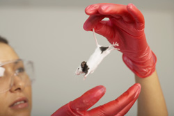
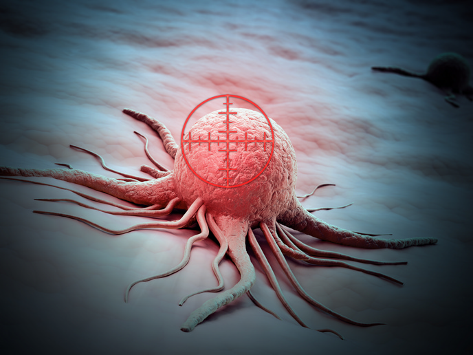
Contemporary radiation therapy for cancer involves the application of complex radiation fields to eradicate the tumour while sparing surrounding healthy tissue. Unfortunately, systems for irradiation of small animals are not so selective and are only able to administer large radiation fields which cannot be modulated with respect to space and time. The net effect of this is that the results of animal experiments cannot be applied to human radiotherapy. During studies of radiation effects on tumours and surrounding cells in small animals, there is therefore a need for development of improved irradiation devices, especially when investigations include the synergistic effects of different cancer treatments. The 'Development of a spatio-temporal-energetic radiation research platform for animals' (Sterrpa) project is developing a versatile small animal irradiation device. The system will be capable of delivering precise radiation fields with modulation available in space and time variables. A small animal precision irradiator/imager, the 'Smart small animal radiation therapy' (Ssmart) device, has been tested and procedures developed for its improved operation. The scientists carried out refinements and tests in three main spheres. Hardware was assessed for irradiation and dose measurements. The Sterrpa team developed treatment planning software whereby animal X-ray computed tomography (CT) scans could be converted into calculation phantoms. A new dosimeter was developed and, using Monte Carlo simulations, accurate determinations of dose were calculated. In one gantry rotation a whole animal three-dimensional (3D) cone beam CT image can be constructed. Image quality was improved by newly developed algorithms and CT images were made suitable for dose calculations. Although not part of the original objectives, Sterrpa scientists took the opportunity to develop algorithms to calculate the true received dose by the animal. Differences between the pre-calculated photon fluence (quantity of X-radiation), and that actually measured indicates discrepancies that can be identified. The final part of the project will be devoted to the development of a machine with high accuracy in irradiation precision and imaging quality that can be used for image-guided radiation studies. The result will be radiation studies in conjunction with the synergistic effects of drugs or response enhancers. Applications will include human cancer treatment and the development of novel treatments in clinical practice.