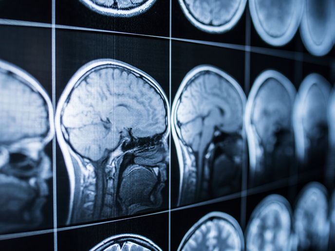
A closer look at the human brain
The EU-funded project ʹComputational neuroimaging: quantitative models of human visual neuronsʹ (PRF MODELS) proposed to bridge this gap by coupling non-invasive human neuroimaging signals with neuronal properties. The researchers used a new computational neuroimaging method which measures human neuronal population properties that are close to those derived from invasive animal experiments. The utility of this method extends from fundamental to clinical neuroscience, as is supported by observations in both rare and common disorders that cause visual impairment. The PRF MODELS team first wanted their computation measurements to replicate well-established animal experiments and human behavioural data. They then validated their measurements with predictions derived from an established theoretical framework. Finally, they aimed to build more complex models of neuronal properties under natural viewing conditions. Neuropsychological evaluation of two stroke patients revealed specific deficits in motion perception. These results provided evidence for several distinct mechanisms to process motion in the human visual system. The project also provided evidence for both stability and plasticity in a rare human congenital disorder known as achiasma. The PRF MODELS method can be applied to basic and applied neuroscience, as indicated by initial studies of achiasma and more common disorders like macular degeneration.