Investigating blood flow following a stroke
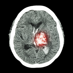
According to the European Heart Network, stroke is the second most common cause of death in Europe and the leading cause of long-term disabilities. An ageing European population means that the human and economic costs of stroke are likely to increase significantly.To address this problem, thie 'Neurovascular coupling in stroke--the brain microvasculature as a target for prevention of ischemic brain damage' (NEUVASCHEMIA) project looked at a key mechanism--regulating blood circulation in the days following a stroke. Earlier research had shown that pial arteries in the brain stimulate some receptors that inhibit blood flow after a stroke, leading to decreased cerebral circulation. However, pial arteries contribute only half of the vascular resistance in the brain. The other half comes from branches called brain parenchymal arterioles. When the brain is busy with a cognitive task, the local brain cells, called astrocytes, signal that increased blood flow is needed through extensions called endfeet. This process, called neurovascular coupling, serves to increase blood flow. Understanding the mechanisms controlling neurovascular coupling was the focus of this research.Experiments involved comparing neurovascular coupling in acute brain slices from sham-operated rats (the control group) and rats induced with global cerebral ischemia, or restricted blood circulation. To initiate neurovascular coupling, brain slices loaded with the calcium indicator Fluo-4 were stimulated through electrical fields. Researchers then measured the calcium signals in the astrocytic endfeet and diameter changes of the parenchymal arterioles in response to neuronal stimulation. The results showed that neurovascular coupling no longer occurred in rats subjected to global cerebral ischemia. To further understand why, researchers analysed each part of the process. They found that calcium signals were unchanged, but there was reduced function of potassium channels in the small muscle layer of the cerebral parenchymal arterioles. These channels play a key role in supplying brain tissue with blood. This discovery deepened understanding of the molecular mechanisms responsible for preventing neurovascular coupling. It could lead to new therapeutic strategies targeting the cerebrovascular system.