Full-service surgical imaging
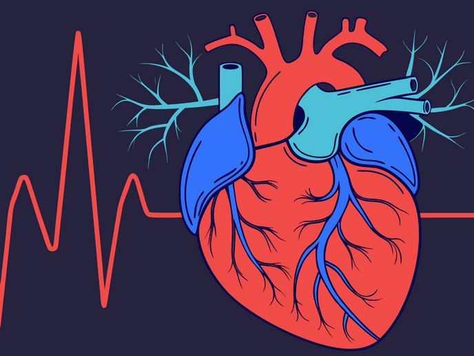
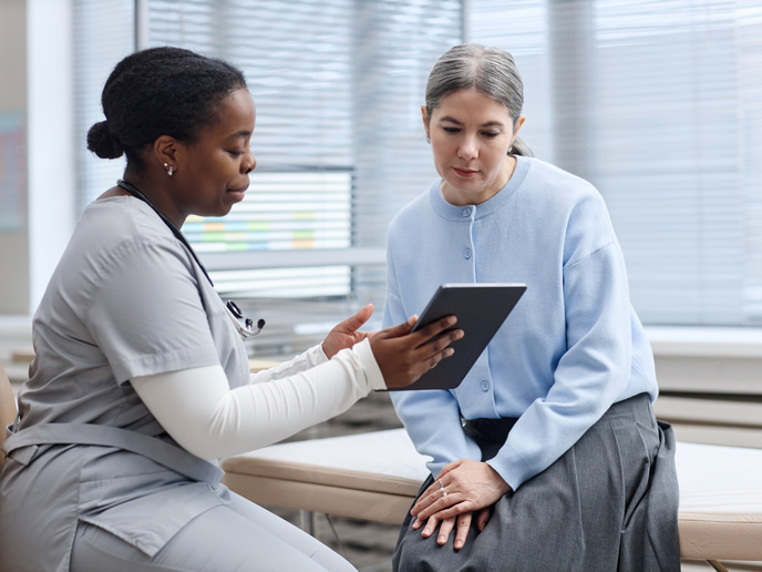
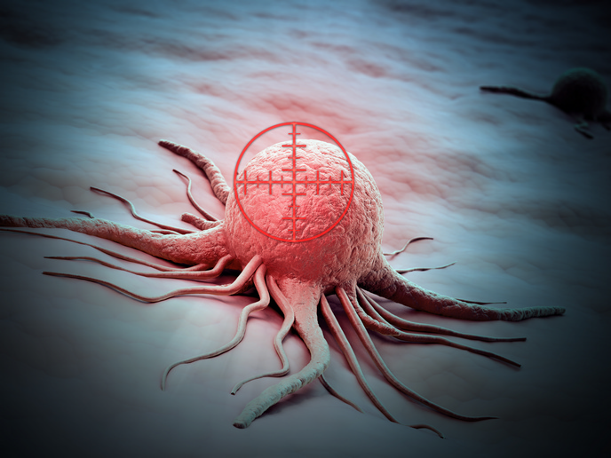
The use of MRI during surgery or other interventions is limited due to incompatibility with devices such as cannula, guided wires or catheters. It is also not compatible with ultrasound and newly developed biophotonic-imaging probes. To exploit the full potential of MRI for real-time image guidance during surgical interventions, a large multi-disciplinary consortium launched the project 'Integrated interventional imaging operating system' (IIIOS). IIIOS combined the expertise of two industrial imaging partners, six university hospitals and two biomedical technology societies. In addition, funding facilitated recruitment of 16 early stage researchers and six experienced researchers with expertise ranging from device development and medical physics to anaesthesiology. Their ambitious goal was integration of interventional and surgical devices with ultrasound and biophotonics-based imaging, MRI, computed tomography and positron emission tomography. Achieving it required intense technology and procedure development. This was supported by monthly technical web-based conferences, training events and two international summer schools. The team engaged in extensive software development to deliver models, simulations, and image analysis and tracking algorithms. Scientists developed a setup enabling interventional MRI for cardiovascular and percutaneous interventions (accessing inner tissue through a needle puncture of the skin). They created and validated surgical equipment compatible with MRI setup. Among the many devices were cardiovascular implants, biophotonic probes, a steerable guide wire and catheter and high-resolution electrocardiography and electroencephalography amplifiers. In the end, the team completed a clinical study of 27 patients with liver carcinomas employing MR-angiography procedures to visualise the organ and vessels. The highly prolific IIIOS project enabled publication of four book chapters, 23 scientific papers, 28 conference proceedings and 71 conference posters and presentations. Two patent applications have already been filed. Combining the most advanced imaging techniques available into a single imaging platform for diagnosis and intervention has pushed the frontiers of medicine. Partners now have in their hands state – of – the – art technology with 22 scientists expertly trained in that technology. Hospitals and patients will feel the impact of this dedicated work for a long time to come.