The molecular basis of cell polarity
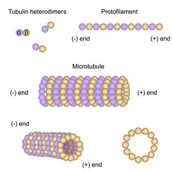
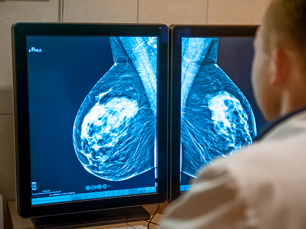
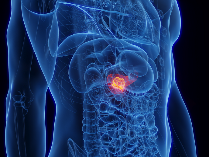
Cells have an intrinsic capacity to polarise their structure and function. This means that following an extracellular cue, they asymmetrically distribute elements of the cytoskeleton and membrane system, leading to polarised membrane traffic. This property facilitates many biological processes including tissue development, neural transmission and immune responses. Central to cell polarity is the microtubule cytoskeleton. Accumulating evidence indicates that plasma membrane events, including receptor activation, integrin signalling and recruitment of certain factors are necessary for microtubule stabilisation. However, the regulatory mechanism of microtubule stabilisation and polarity remain elusive. The scope of the EU-funded CELL POLARITY (Role of microtubule polarity and polarized membrane traffic in directed cell migration) project was to delineate these molecular mechanisms and identify factors central to directed trafficking. For this purpose, they established a wound-edge fibroblast system and used high-/super-resolution imaging methods to quantify microtubule changes after knockdown of specific factors. Research results indicated that two evolutionarily conserved polarity proteins, Numb and Par3, had opposing roles in microtubule stabilisation. The endocytic adaptor Numb acted as a novel suppressor of microtubule stability, while the polarity scaffolding protein Par3 was necessary for stabilisation. This Numb/Par3 interplay might finetune the amount of stable microtubules, thereby regulating the speed or persistency of directed cell migration. In addition, scientists expressed fluorescent Par3 to visualise its spatiotemporal behaviour in migrating cells. They identified two distinct pools of Par3, one static at cell-cell contacts, and one dynamic at the leading edge. Identification of the biological significance of the dynamic pool would help delineate its similarity to actin retrograde flow and microtubule stabilisation. Furthermore, scientists used novel fluorescence-based super-resolution imaging methods such as structured illumination microscopy, stimulated emission depletion and direct stochastic optical reconstruction microscopy. These provided enhanced resolution over conventional methods to analyse the spatiotemporal organisation of sub-cellular proteins and organelles during cell polarity. Overall, the CELL POLARITY study provided novel mechanistic links between integrin endocytosis, phosphoinositide switching and microtubule stability. Given the role of many of these factors in diseases such as cancer and neurological dysfunction, the project findings provide new targets for therapeutic intervention.