

EMBL scientists achieve breakthrough in microscope technology
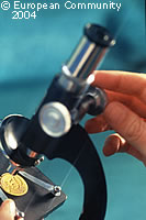
Scientists at the European Molecular Biology Laboratory (EMBL) have come up with a revolutionary new microscope which, for the first time, allows an observer to study living specimens from different angles under real conditions. The device has been named SPIM (selective plane illumination microscopy), and EMBL believes that it is certain to become a standard fixture in biology laboratories in the future. 'Over the years we've seen current microscopes falling short of what the scientists need,' explains Ernst Stelzer, whose EMBL group developed SPIM. 'We designed SPIM with EMBL biologists to make sure that it was completely suited to their needs. This microscope is easy to build, is about one third the cost of current technologies, and gives scientists improved resolution by a factor of about five.' SPIM enables scientists to view relatively large samples in a medium that mimics real conditions, rather than destroying the sample by slicing it up to fix it into slides, which is the traditional method. The new instrument then shines a very thin slice of light through the sample, moving the specimen through the light to capture images from each layer. Not only does SPIM give a sharper image, but the whole specimen can also continue living and growing as it is being viewed, something which is impossible using current microscopes. The procedure is also very fast, providing detailed images in minutes, and by rotating the sample and scanning it repeatedly, the combination of views yields an unprecedented three-dimensional (3D) image. Dr Stelzer concluded: 'It is extremely valuable - especially for studying developmental biology and for viewing 3D cell cultures. With SPIM, scientists can now capture images that would have been otherwise impossible. It enables completely new applications in scientific research.'