Cracking the code of vision
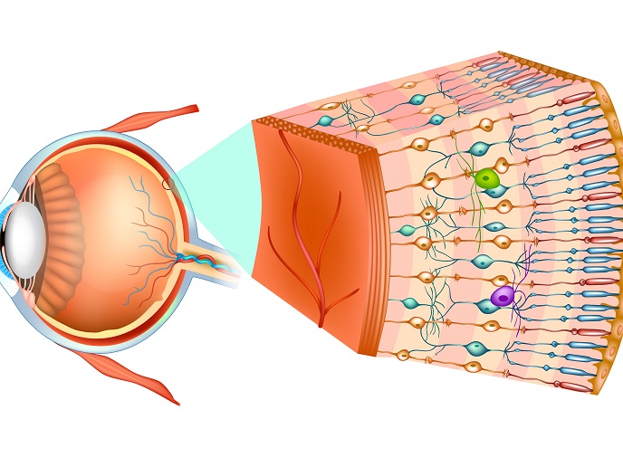
The retina is a thin sheet of neural tissue at the back of the eye. It contains a layer of specialised cells called photoreceptors that capture the incoming light, transform it into electrical signals, and pass them through a complex network of neurons that include bipolar and ganglion cells. These refine and amplify the signals before they are sent to the brain via the optic nerve. However, there are many aspects about our sense of vision that remain unclear, such as how these electrical signals represent visual information of natural scenes with real-world objects, motion, and alterations in illumination conditions.
Insight into neuronal circuit of the retina
The key objective of the CODE4Vision(opens in new window) project, funded by the European Research Council, was to better understand visual signal processing and develop therapeutic strategies for treating diseased retinas. Researchers applied a new method for recording neuronal signals in retinal cells alongside a powerful data analysis technique. Collectively, these helped to visualise an important part of the retinal circuitry: the layout of connections between the retina’s layer of output neurons and the preceding layer of excitatory inputs. The new insights about the connections in the retina led to the development of new mathematical models(opens in new window) to describe how the retina encodes natural visual scenes. “We now better understand why distinctive types of retinal output neurons respond differently to the same scene and what their roles are in vision,” explains principal investigator Tim Gollisch.
Treatment of retina conditions
Retinitis pigmentosa(opens in new window) is a disease of the eye associated with degeneration of photoreceptors, which affects millions of individuals worldwide. As no cure is available, considerable efforts have been made for restoring vision. A promising strategy involves the insertion of light-sensitive proteins into surviving retinal neurons, turning them into artificial light sensors, an approach known as optogenetics. Evidence indicates that normal light from the environment does not lead to proper responses in these artificial light sensors. Therefore, CODE4Vision researchers calibrated the visual stimulation apparatus to achieve the necessary levels of bright light that trigger the implanted artificial light sensors within the retina. Through the utilisation of CODE4Vision analysis and mathematical models, researchers were able to refine the modifications for the projection, resulting in an optimised application of optogenetic therapy for retinitis pigmentosa. This was validated in a mouse model of the condition where improved responses were observed by artificial stimulation. “By making the artificial stimulation of the retina more natural, we hope that it becomes easier for the brain to understand the visual scene, leading to a better restoration of visual sensations,” emphasises Gollisch.
The future of vision restoration therapies
Collectively, CODE4Vision findings provide a better understanding of how the neural circuit of the retina is orchestrated and how it processes natural visual scenes. “Investigation of the encoding of natural stimuli by the retina is an ongoing process, and we plan to continuously update and extend our models,” outlines Gollisch. The team is also interested to unveil how contextual information, like the average light intensity or the contrast in the current environment, influences this encoding. Moreover, to improve the therapeutic approach of artificial stimulation, they will test different types of light sensitive molecules to identify the most promising candidates for future clinical exploitation.