New techniques herald personalised approach to eye diagnostics
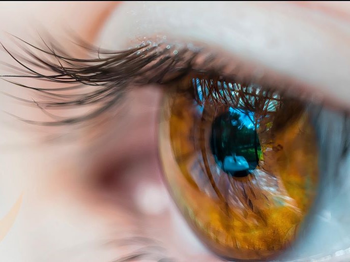
Eye specialists currently use parameters such as corneal topography or examining the shape of the cornea to screen for keratoconus(opens in new window), a disease of the eye. However this will only detect the problem once damage to the cornea is already happening. Fresh approaches, and a better understanding of corneal properties, are needed to tackle this and other serious eye conditions.
Revealing eye biomechanics
The EU-funded IMCUSTOMEYE(opens in new window) project set out to develop a non-invasive imaging device, capable of characterising the biomechanics of the cornea, in order to improve both screening and treatment of several diseases. “Measuring corneal biomechanics is an unmet need. To date there is no commercial instrument that provides a map of corneal mechanical properties,” says Susana Marcos, project coordinator and director of the Visual Optics and Biophotonics Lab(opens in new window) at the Spanish National Research Council(opens in new window). The team used nanosensitive optical coherence tomography(opens in new window) (OCT), combined with a non-contact corneal stimulus, to deliver new imaging techniques which were validated in the lab and demonstrated in the clinic. The system provides an unobstructed view of corneal deformation from multiple angles. It can also capture three-dimensional OCT-based corneal topography, thereby generating tomographic and biomechanical measurements with just one tool. Biomechanical properties are obtained from coupling the imaging device and the deforming stimulus. This causes macro-deformations (air puff), modulated micro-deformations (sound) or nanometre-amplitude laterally propagating waves (ultrasound) in the cornea. The team came up with a way of translating the deformation amplitudes, resonance frequencies and propagating wave speeds into biomarkers for measuring corneal health. This approach could be useful for producing early diagnostics tools for screening.
Baseline for healthy corneas and early diagnosis
These new techniques were incorporated into compact prototypes and used to establish a baseline for corneal biomechanics in a healthy population. The team used them to conduct clinical studies in patients with keratoconus, others pre- and post-LASIK surgery and, finally, a third group of patients who had undergone intracorneal ring segment surgery and corneal cross-linking. The resulting data was used to produce corneal biomechanical maps and to identify biomarkers for corneal abnormality. “We are able to identify keratoconus based on the comparison of the biomarkers with those of a normal population, helping diagnosis and identifying patients with a potential risk of ectasia,” explains Marcos. “The device is sensitive – able to detect keratoconus at a very early stage, prior to any other symptoms,” she adds. But that is not all. Project data was also used to simulate corneal response to different pressures.
Personalised patient data
Ophthalmologists rely on standard measurements to estimate interocular pressure (IOP) or the fluid pressure of the eye. High eye pressure is the biggest risk factor for glaucoma, so the ability to accurately measure each patient’s IOP could significantly improve outcomes. “Corneal mechanical properties affect standard IOP measurements,” notes Marcos. Being able to measure these in every patient will be relevant for accurate IOP and therefore diagnosing glaucoma. IMCUSTOMEYE’s market research indicates potential demand for a more sophisticated device capable of mapping corneal biomechanics and topography – which could help with planning surgical interventions – and a second, cheaper device for use in screening for disease and IOP measurement.