Innovative imaging captures how ageing and disease impact bones
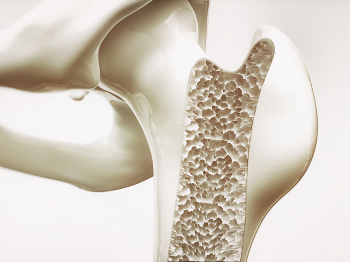
Osteoporosis(opens in new window) is a condition where bones become weaker and more fragile as their internal structure deteriorates, resulting in an increased risk of bone fracture, even from minor falls or routine movements. “While often associated with ageing and hormonal changes, osteoporosis can also be caused by chronic inflammation, which disrupts the balance between bone formation and bone resorption, leading to accelerated bone loss,” says Georg Schett coordinator of the 4-D nanoSCOPE(opens in new window) project, which was funded by the European Research Council(opens in new window). This inflammation-driven form of osteoporosis is particularly relevant for patients with chronic inflammatory or autoimmune conditions(opens in new window) yet is often under-recognised, a situation 4-D nanoSCOPE set out to rectify. The project sought to add to the knowledge base about how inflammation affects bone structure across different spatial scales and over time, and how these structural changes translate into reduced bone strength. The project achieved this thanks to the development of a new generation of imaging and analysis tools. “Alongside characterising and analysing bone structure across different scales, our approach can correlate structural features with biological processes and mechanical properties, providing insights into bone strength and the influence of diseases and treatments,” notes Schett from University Hospital Erlangen(opens in new window), the project host.
Developing the integrated imagining workshop
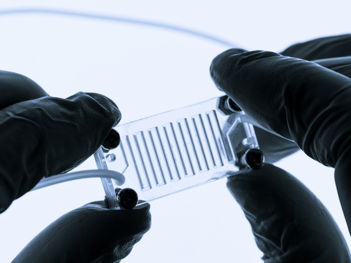
Prior to 4-D nanoSCOPE existing bone imaging methods were either too limited in resolution, restricted to specific scales or unable to capture changes dynamically over time. Consequently, it had not been possible to observe bone modelling in a truly 4D way – three spatial dimensions plus time, especially under realistic biological conditions. 4-D nanoSCOPE developed a solution combining advanced X-ray nanoscopy(opens in new window), complementary imaging approaches and AI–based data analysis, merged into one integrated workflow. Once bone samples are generated they are imaged in 3D using X-ray microscopy and then analysed quantitatively by AI tools. These identify microstructures quantifying their properties to track changes over time. Unlike conventional approaches relying on large-scale facilities, this system is operational in standard laboratory environments, opening up access to high-resolution imaging. Additional technical innovations included: a high-speed, highly sensitive detector allowing rapid image acquisition while reducing radiation exposure and sample holder and imaging protocols ensuring that samples can be positioned and imaged consistently.
Supporting EU health and digital innovation priorities
The project carried out a series of experimental studies and computational analyses. “Our results demonstrate that bone can now be studied in a more integrated, quantitative and time-resolved way than before. And importantly, we were able to compare different causes of bone loss, including inflammation, ageing and hormonal changes,” explains Schett. By improving diagnosis, 4-D nanoSCOPE points to a future of more targeted preventative strategies and treatments, improving quality of life for affected individuals and supporting more sustainable healthcare systems. The team’s next steps focus on further developing and applying the project’s technologies and methods. “We aim to get closer to clinical use, for example by integrating our approaches into research that supports diagnosis, treatment monitoring and therapy development,” adds Schett. In parallel, the project’s workflows and datasets will be shared within the scientific community to enable follow-up studies and broader application, opening up new opportunities for interdisciplinary collaboration in fields such as musculoskeletal research, materials science and biomedical engineering.