The next generation of brain imaging

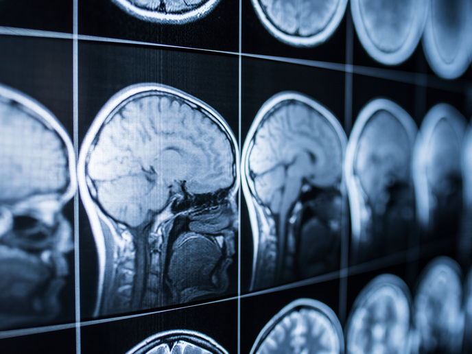
Neurological diseases often begin with subtle changes inside individual cells, long before conventional brain scans reveal visible damage. This delay can make early diagnosis difficult and limit clinicians’ ability to distinguish between different disease processes or monitor how patients respond to treatment. Conventional MRI is an indispensable clinical tool for identifying where damage has occurred in the brain, but it cannot always explain what biological processes are responsible. Neurodegenerative disorders such as Alzheimer’s disease, neuroinflammatory conditions such as multiple sclerosis and brain tumours all affect different cell types and trigger distinct pathological mechanisms that often appear similar on standard scans.
Moving beyond conventional MRI
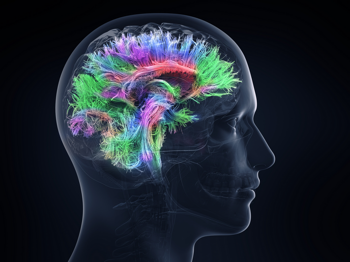
The ERC-funded C-MORPH(opens in new window) project set out to develop advanced MRI techniques capable of detecting cell-specific changes in the living human brain and spinal cord, opening new possibilities for personalised neurological care. The goal was to move beyond the production of anatomical images, but rather provide information about cell density, morphology, intracellular organisation, metabolism and tissue composition. To achieve this, researchers combined advanced diffusion MRI(opens in new window), MR spectroscopy, ultra-high-field imaging and computational modelling. “We wanted to identify cell-specific MRI biomarkers associated with the underlying cellular changes occurring in living tissue,” explains principal investigator Henrik Lundell.
New imaging methods reveal hidden biology
Researchers created new tools for characterising structural features of cells, with a particular focus on neuronal tissue. Their work demonstrated how novel contrast mechanisms(opens in new window) can simultaneously distinguish the size and shape of microscopic structures on the micrometre scale. The project showed that advanced diffusion methods can better characterise tissue microstructure while reducing interference from factors such as fibre orientation. Approaches were also developed for distinguishing inflammatory activity from permanent tissue damage, providing valuable insight into disease progression and treatment response. “We have developed advanced MRI methods that exploit the motion of water and metabolites to probe biological processes occurring at the cellular scale,” states Lundell. By combining information from water diffusion(opens in new window), metabolite mobility and tissue composition, researchers can investigate changes in specific cell populations before large-scale structural damage becomes apparent. Moreover, the project uncovered an entirely new imaging phenomenon known as spectral anisotropy. Initially described theoretically and through computer simulations, the effect has now been demonstrated experimentally for the first time.
Towards clinical translation
Although many of the techniques have reached technical maturity, further validation in larger patient cohorts is required before routine clinical implementation. According to Lundell, some quantitative and advanced diffusion MRI methods could enter specialised clinical centres within the next five years as scanner hardware and software continue to evolve. More complex metabolic imaging approaches will require additional multi-centre validation and standardisation before widespread adoption. The project also continues to explore the clinical potential of spectral anisotropy and other newly developed biomarkers in patient studies. Beyond neurological disease, the methods are expected to provide valuable insights into healthy brain development, ageing, learning and the influence of lifestyle on brain health.