

Osteoporosis warning from dental radiographs
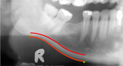
Progressive reduction of bone mineral density (BMD) or osteoporosis, is a silent process which is quite often only evident on fracture of hip, wrist or vertebral column. The patient is then faced with a long period of rehabilitation when a substantial period of time may be lost from work or they can suffer a severe reduction in their life quality. Advanced notice of the onset of osteoporosis would mean that the patient could obtain pharmaceutical intervention or adopt appropriate lifestyle changes. Researchers based at the University of Manchester as part of the OSTEODENT project, developed software to analyse dental radiographs of the lower jawbone. Many millions of these panoramic dental radiographs are taken by European dentists each year. The project had recruited 650 peri-menopausal women and identified those with osteoporosis, normal and reduced bone mineral density, measured by dual energy X-ray absorptiometry (DXA) of the hip and spine. Routine manual estimations of cortical width on radiographs by dentists show variability within and between operators, as well as being quite time consuming. The aim of the project was to develop automatic computer software to analyse the radiographs and thereby minimise reproducibility error and maximize diagnostic accuracy of osteoporosis. The scientists trained an Active Shape Model of the cortex using radiographs from another large dataset. The software then automatically measured the cortical width between two points on the lower jaw. A highly significant correlation was found between cortical width and BMD Furthermore, using receiver operating characteristic (ROC) analysis, the results were clinically useful with good sensitivity and specificity values. Advantages are that the use of the software involves little or no user interaction and its diagnostic validity is high. Applications are twofold. The accurate advanced diagnosis of osteoporosis using a fairly routine visit to the dentist is the most obvious. However, assessment of jaw bone density may be commercial use when planning dental implants Using this analytical radiographic software, patients could be identified as to their suitability for dental implant procedures. Figure caption: Automatic measurement of cortical thickness on a radiograph