Novel endoscopic tool advances cancer imaging
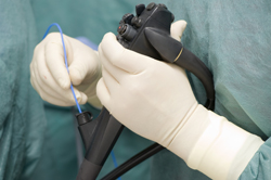
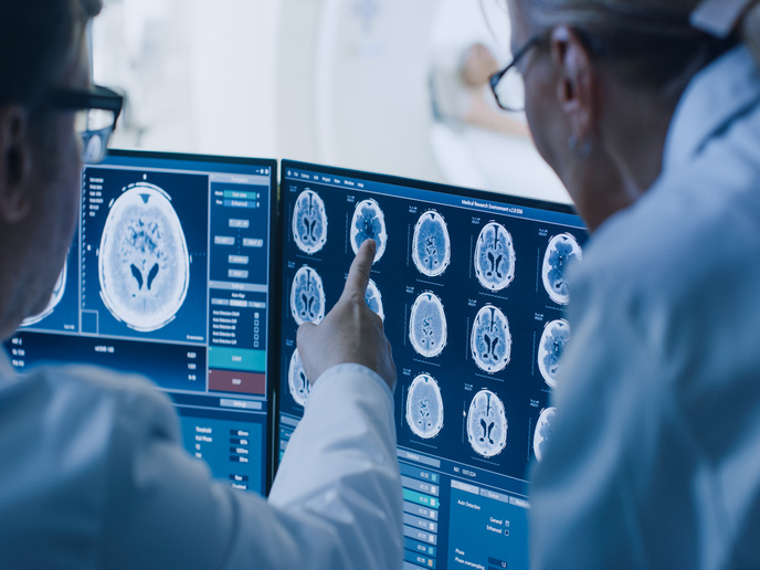
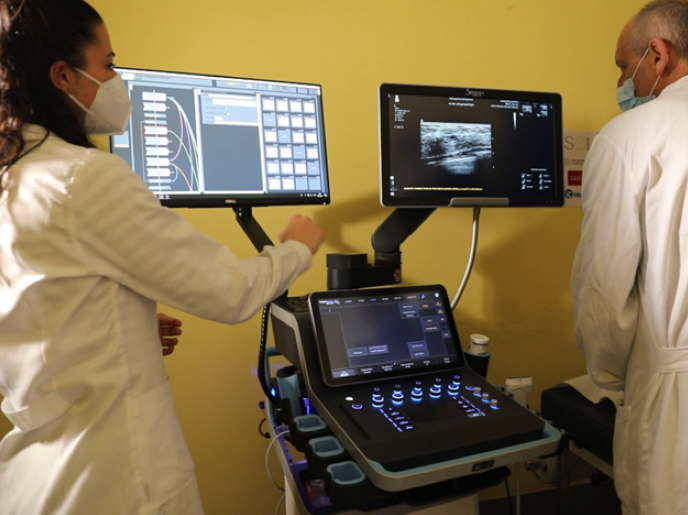
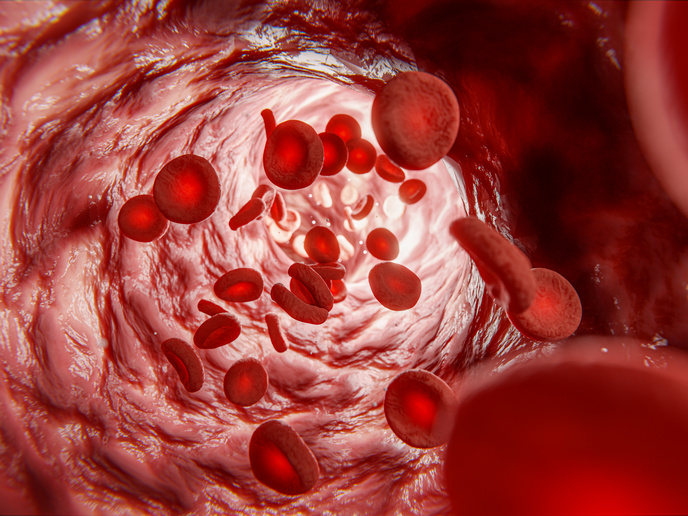
Certain cancers (pancreatic and prostatic, for instance) are asymptomatic, commonly having poor prognosis and calling for invasive surgical treatment. Less invasive and more precise microsurgical interventions combined with new diagnosis protocols based on specific biomarkers could improve the prognosis in such malignancies. To this aim, the EU-funded Endotofpet-US project proposes to build a bimodal positron emission tomography and ultrasound (PET-US) endoscopic probe in a miniaturised system. To test the procedure, partners are launching a pilot clinical study focusing on pancreatic cancer, following preclinical feasibility assessment in pigs. On the back of recent technological advances in photodetectors and scintillators, scientists have constructed a bimodal endoscopic probe to assist image-guided diagnosis and minimally invasive surgery. This novel piece of equipment boasts spatial resolution down to 1 millimetre, and a 100-times-higher sensitivity than whole-body PET scanners. Optimised LSO type crystals are being used to achieve a timing resolution of 200ps. Among the project’s challenges is to integrate all the components in a very compact detector head with an efficient tracking system. A prototype of a coupling optics plate has been produced and characterised based on the design of the photosensor. With regard to the front-end electronics for the external plate and the data acquisition system, project partners are exploiting the time over threshold (TOT) approach using a special low-noise NINO chip developed at European Organization for Nuclear Research (CERN). In comparison to existing endoscopic imaging, which provides structural and very limited functional information, the ENDOTOFPET-US probe will produce high-sensitivity functional information during endoscopy. It can thus allow for highly specific PET biomarkers accumulating in very small structures to be easily distinguished from the background. This would facilitate analysis beyond the walls of the examined lumen, while at the same time providing invaluable data on specific biomarkers.