Less harmful medical imaging techniques
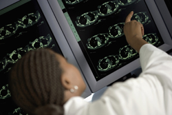
New diagnostic procedures such as computerised tomography (CT), positron emission tomography (PET) and single photon emission computerised tomography (SPECT) entail the introduction of radiopharmaceuticals to patients. This results in increased radiation exposure of healthy tissues as well. The EU-funded 'Minimizing activity and dose with enhanced image quality by radiopharmaceutical administrations' (Madeira) project was designed to address this problem. By performing biokinetic investigations and modelling, partners obtained important knowledge of the radiation exposure associated with each of the most widely used medical imaging techniques. At the same time, the Madeira project intended to develop new add-on hardware (PROBE system) in order to enhance spatial resolution of existing PET systems and obtain three-dimensional (3D) information. To test and compare the different approaches, a specialised software tool was created enabling image processing. Further developments enabled scientists to calculate relevant dose distributions in the body and to optimise signal-to-noise ratios in order to reduce the radioactivity required to obtain a good image. Collectively, the methods developed during the Madeira study were intended for personalised medicine. The ultimate goal was to assess and optimise the radiation required for each patient to obtain successful diagnostic information with a particular nuclear medical imaging technique. Training courses and conferences ensured appropriate dissemination of the project's results. Partners are hopeful that their methods have the potential to reduce the administered radioactivity at least a factor of three without compromising the sensitivity or resolution of the imaging techniques.