Better technology to track diseases
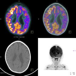
Positron emission tomography (PET) is an imaging technique that uses a special camera and a 'tracer' molecule to visualise organs and look for disease. The tracer is prepared by attaching a radioactive atom, most often fluorine-18 (18F), to a molecule used by the body (e.g. glucose) to create a radionuclide. 18F is fairly unstable, with half of its atoms decaying in 110 minutes. This means that the radionuclide must be injected into the body immediately after it is made. In addition, 18F is currently limited to tagging small, simple molecules like glucose. To expand the capabilities of PET to more biomedically interesting, complex molecules, scientists developed new radioactive tracers with EU funding of the project FLUOPET (Late stage fluorination for positron emission tomography applications). Researchers built on recent advances in the field using 19F, the most stable and abundant form of fluorine, to fluorinate more complex molecules. They wanted to adapt the chemical methods developed for 19F to the 18F version used for PET imaging. First though, they developed protocols to fluorinate complex molecules like nerve cell receptors for brain imaging with the standard 19F atom. During these experiments, scientists created a fluorine-containing liquid that that is more useful than its normal gaseous form. This patent-pending invention will allow chemists to prepare fluorinated compounds more easily. By the project's end, FLUOPET made two new PET tracers for imaging proteins and receptors implicated in cancer and neurodegenerative disorders. This work will greatly advance the capabilities of an already powerful non-invasive technique to diagnose and monitor important diseases.