

Scientists piece together myosin-actin puzzle
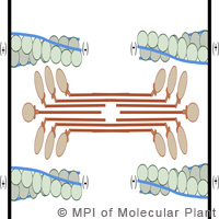
Researchers from Germany and the United States have succeeded in uncovering how myosin and actin filaments work together to regulate muscle and other movement processes. They provided an image of the proteins tropomyosin and troponin that control how myosin binds to actin. The findings of this study could help researchers establish how genetically determined modifications affect the actin-myosin-tropomyosin complex in some types of hereditary heart disease. Led by Stefan Raunser and Elmar Behrmann from the Max Planck Institute of Molecular Physiology in Germany, the team generated an accurate image of the actin-myosin-tropomyosin complex to 0.8 nanometres by using sophisticated electron microscopy techniques. The image represented a resolution of less than one-millionth of a millimetre. This was never been done before. Thanks to this study, researchers could correctly identify the location of proteins within the complex and analyse muscle contraction processes. The sarcomere, which is a muscle's basic functional unit, comprises actin, myosin and tropomyosin proteins. The contraction of a muscle means the myosin must slide along filamentous actin molecules. Tropomyosin, along with troponin, controls muscle contraction by regulating when myosin binds to actin, according to the researchers. The binding site for myosin on the actin filament is blocked by tropomyosin and troponin in the resting state, they said. 'The myosin head is at a 90-degree position,' the researchers said. 'Only after an influx of calcium, which docks onto the regulating proteins, is the binding site on the actin filament exposed. The myosin head docks onto this site, changes its conformation and bends in an articulated manner, thereby pulling the actin along with it.' The filaments slide over each other, leading to a shorter sarcomere and muscle contraction. 'This is an important step in understanding the interplay between the individual proteins within the functional structures of muscle,' said Dr Raunser. 'We have, so to speak, drawn a map for biochemists. Our findings will make it easier for them to understand the processes and sequences of events taking place in muscles.' The results will also benefit medical specialists, who seek to provide insight into the malfunctions within the heart, which are usually linked to point mutations. 'Finding the exact location of the mutations is fundamental to developing treatments for such heart diseases,' said Dr Raunser. Researchers from Hannover Medical School and Ruhr-Universität Bochum in Germany and the University of Texas in the United States contributed to this study.For more information, please visit:Max Planck Institute of Molecular Physiology:http://www.mpi-dortmund.mpg.de/english/Start/Welcome/index.htmlHannover Medical School:http://www.mh-hannover.de/index.php?L=1
Countries
Germany