Improving breast cancer diagnostics with robotics

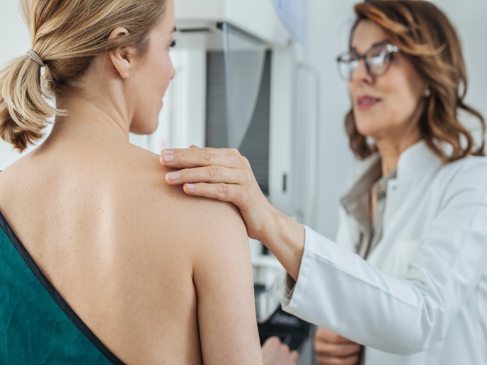
Breast cancer is among the deadliest diseases in the world, affecting one in eight women. Early detection is key to treating the condition in time, saving lives and reducing the overall impact of the disease. Improving the tools and procedures used for breast cancer diagnostics plays a major role in this effort. The EU-funded MURAB project(opens in new window) has taken a decisive step towards next-generation diagnostics. “Our new robotics-assisted surgical system could make a big difference for patients, doctors and healthcare systems,” explains Stefano Stramigioli, professor of Advanced Robotics at the University of Twente(opens in new window) and MURAB project coordinator. “This is achieved by applying the precision of robotics to the precision of imaging.”
The best of two worlds
Currently, two different procedures exist for detecting anomalies in the breast and carrying out biopsies, each with its own limitations. While ultrasound offers real-time imaging, lower costs and wide availability, it is ineffective for detecting very small lesions. Magnetic resonance imaging (MRI), on the other hand, offers high-quality visualisation and better detection, but the absence of real-time imaging makes MRI-assisted biopsies a difficult, lengthy and costly procedure. MURAB offers a solution to this dilemma while reducing the risk of human error. Stramigioli explains: “The new procedure would start with an MRI scan of just up to 15 minutes revealing the smallest lesions, followed by a robot-assisted ultrasound examination and biopsy.”
High precision
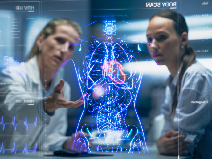
Under the new system, a smart robot will combine the data from the MRI with an ultrasound scan of the breast, enabling high-precision localisation of the lesion. Based on this information, the robotic arm can then steer the needle – which is manually inserted by the radiologist – to the exact location where the biopsy needs to be performed. “This workflow not only reduces discomfort and uncertainty for the patient, but also effectively eases the workload for clinicians and hospitals, potentially enabling them to handle more cases faster,” Stramigioli says. In addition to its applications in the field of breast cancer diagnostics, the concept could also be used for muscle biopsies.
From diagnostics to cure?
Thanks to the EU funding, the project team has now delivered the proof of concept, paving the way for clinical trials which will take the system one step closer to becoming available to patients and carers. By combining expertise from the fields of robotics, imaging, ultrasound and breast oncology and involving research as well as business partners, the consortium has laid a solid foundation for turning this concept into an actual product. “With the proper funding and the right market conditions, it is possible to bring the new system to the market within around 5 years,” adds Stramigioli, cautioning that further large-scale investments will be required to get there. By upgrading the needle component using optical fibre, the concept could evolve to allow for carrying out not only biopsies, but also ablations. “With this future development, a woman could go to the hospital for a routine check-up and come out a few hours later cured from a cancer she didn’t know she had,” he concludes.