Searching the eyes for telltale signs of Alzheimer’s

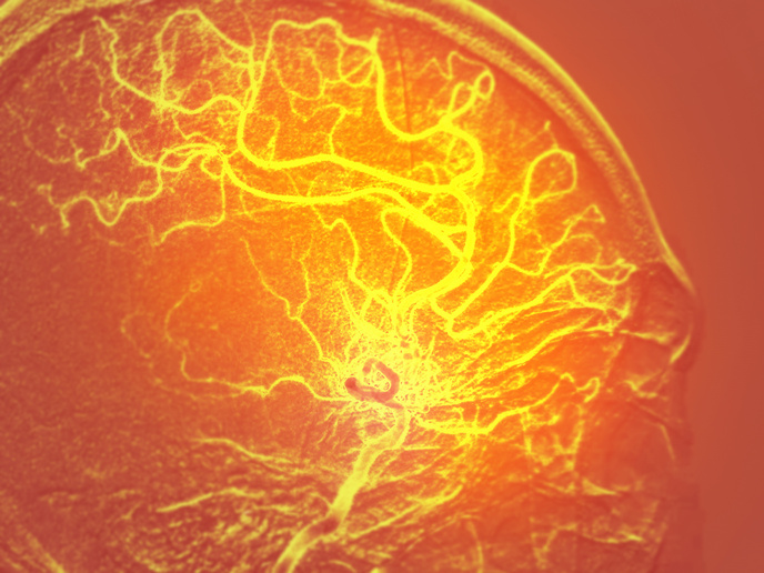
Neurodegenerative diseases such as Alzheimer’s affect 10 million people in the European region, and that number is expected to double by 2030(opens in new window). However, diagnosing neurodegenerative diseases remains difficult, and suspected cases can only be conclusively determined by examining the brain post-mortem. One promising avenue of research is to use ophthalmology techniques to look for changes in the eye that could indicate the presence of neurodegenerative disease. This was the object of the EU-funded OPTIMALZ(opens in new window) project. “Things going on in the brain may also exhibit in the neural tissue of the eye, as the eye is part of the central nervous system,” explains project coordinator Bernhard Baumann(opens in new window). “So these can be assessed from the outside, in features which you can see directly.” The technology under development at Baumann’s lab at the Medical University of Vienna(opens in new window) is based on multifunctional optical coherence tomography (OCT). This non-invasive technique is analogous to ultrasound, except that it uses light instead of acoustic waves. OCT allows ophthalmologists to obtain detailed three-dimensional images of the microstructures in the retina.
Visualising pathologies
Baumann and his team investigated three applications of OCT, examining the retina, lens and brain tissue of mice, as well as histological samples of brain tissue from humans. They looked for changes to the vascular microstructures and blood flow, and searched for lesions and deposits of amyloid plaques that are characteristic of Alzheimer’s disease, hoping to correlate pathology in the brain with biomarkers in the eyes. When they started, there were some indications that pathologies in the eyes might relate to neurodegenerative diseases. However, says Baumann, a complex picture emerged: “Depending on the mouse models, we sometimes saw changes in the retina, other times we did not. You have to be really careful how to interpret the data.” Nonetheless, Baumann adds, the OCT technology worked very well to visualise pathologies in the eyes and brain, even if their clinical relevance was not always clear: “It’s quite promising. We have good resolution in real time, we can see tiny lesions.”
All eyes on brain tumours
The work was supported by the European Research Council(opens in new window). “This allowed me to hire a group of people, build new systems and software for analysing data, and have some money for publications and conferences,” notes Baumann. “The great thing about this grant is that you have 5 years to test your research hypothesis and make use of the developments to even further explore fields along the line.” The team plans to continue developing the OCT technology. They recently began a collaboration with a pharmaceutical company to use it to screen mouse models. “We’re also working to make our technology useful in analysing brain tumours. It’s exciting to see how far we can push this,” concludes Baumann.