Replacing liver biopsies with non-invasive techniques
Metabolic dysfunction-associated steatotic liver disease (MASLD), formerly known as non-alcoholic fatty liver disease (NAFLD), affects around one in three people worldwide. It occurs when fat accumulates in the liver. “For most of us, fat accumulation is all that happens,” explains LITMUS(opens in new window) project coordinator Quentin Anstee from Newcastle University(opens in new window) in the United Kingdom. “But in one out of 10 cases, that fat causes inflammation in the liver. And, if that inflammation persists, about one in 10 of those cases develop scarring of the liver that progresses to cirrhosis or liver cancer, even if they don’t drink alcohol. Fundamentally, MASLD is driven by being overweight or having diabetes.”
Non-invasive techniques to replace biopsies
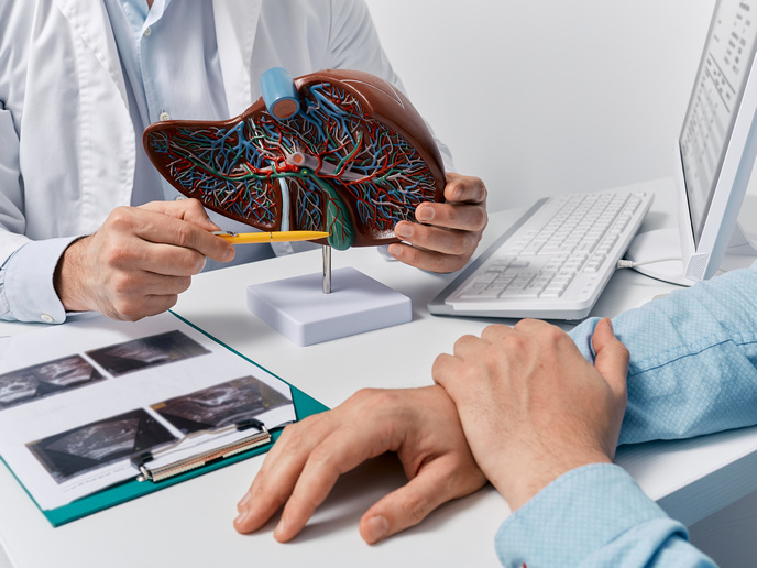
Up until now, the best and indeed only way to accurately diagnose MASLD and assess liver inflammation was to carry out a biopsy. This invasive procedure requires a needle that pierces the liver to collect a sample. The procedure is not without risk and can be an unpleasant experience for patients. The EU and industry-funded LITMUS project was launched to investigate whether non-invasive techniques could replace biopsies. This challenge was addressed in three stages. “First, we carried out a series of systematic reviews of all the existing data currently available on biomarkers,” says Anstee. “We put all this data together, to give ourselves an independent and objective view.” Next, the team pulled together data and biological samples previously gathered in research projects on patients from across Europe. Using these, LITMUS systematically measured multiple biomarkers and conducted a comparative analysis that was published(opens in new window) in ‘The Lancet – Gastroenterology and Hepatology’ last year. The third stage involved recruiting a new cohort of about 2 500 liver biopsy patients. The team collected huge amounts of clinical data, as well as blood samples and MRI scans. A comprehensive biomarker analysis was then carried out. This work is currently being finalised.
New methods for finding biomarkers
Taken together, these three steps enabled LITMUS to collect high-quality data on previously identified biomarkers, and to identify new ones. The project team also conducted large-scale multi-omics studies, using techniques such as machine learning to see if new biomarkers could be identified more effectively. “We now have a really robust understanding of how biomarkers actually perform,” adds Anstee. “We were also able to identify new signals that could be future biomarkers, and we’ve advanced some biomarkers towards regulatory qualification. I’m incredibly proud of what my colleagues have achieved.”
Bringing biomarkers into clinical settings
Anstee recognises that more work still needs to be done to bring biomarkers into clinical settings and ultimately replace invasive biopsies. He identifies the need for a longitudinal follow-up study on patients. This could help clinical researchers move from the diagnostic use of biomarkers, to using biomarkers to monitor patients over time and assess treatment response. “The data generated through LITMUS is already influencing clinical guidelines,” he notes. “This is above and beyond what we set out to do. For example, we are seeing industry partners from the project start to adopt non-invasive solutions as part of their drug development pipelines.” This project has therefore been critical in moving patient care away from invasive liver biopsies as routine practice. “When a third of the population has this condition, and we need to work out who needs care, this is utterly essential,” says Anstee.


