Reliable emergence of order from apparent disorder in the developing retina

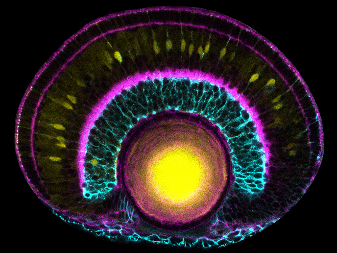
Most neurons are born far from their final destinations. To build a functioning nervous system, these cells must travel through tissue and form functioning connections on arrival. In the retina and other developing brain regions, this includes arranging themselves into distinct layers, a process called neuronal lamination. Little was known about how this happens in the retina. Most research had focused on the cortex, looking mainly at the migrating cells themselves rather than the surrounding tissue. The ambitious ERC-funded project makingtheretina has delivered a step change in understanding of how the retina is formed. Over its six-and-a-half-year duration, makingtheretina used advanced imaging and cross-disciplinary tools to reveal the exquisite dynamic control of neuronal lamination despite seemingly chaotic neuronal ‘traffic’.
Long-term light-sheet imaging of the zebrafish retina
The well-established animal model zebrafish is ideal for observing lamination – it is transparent and small, develops quickly and its retina is relatively accessible compared to other areas of the central nervous system. “Long-term light-sheet microscopy allowed us to watch cell migration and lamination in the zebrafish embryo as it happened,” says principal investigator Caren Norden, previously of the Gulbenkian Institute for Molecular Medicine(opens in new window) and now at the University of Cambridge). This gentle technique illuminates one thin tissue plane at a time, keeping embryos healthy for days. Computer scientists and theorists complemented the experimental work with use of automated analysis software alongside predictive machine learning tools and theoretical models of cell behaviour. “Custom analysis tools allowed us to follow cell movements over time and extract speeds, directions and shape changes, revealing the distinct strategies used by the different retinal neuronal types,” Norden explains. The predictive tools and theoretical models supported iterative hypotheses generation and testing of what drives the movements.
Order emerging from ‘chaotic’ cellular behaviour
“Our studies showed that precise tissue structure emerges from surprisingly messy behaviour. Different cell types move simultaneously using different strategies, often passing each other to reach their final locations,” says Norden. Horizontal cells, for instance, followed unpredictable individual routes rather than fixed paths. They were guided by indirect cell communication – chemical signals from neighbouring cells – rather than physical tissue structure as originally expected. “Most strikingly, some neuronal movement serves to protect tissue maturation. Photoreceptor cells travelled back and forth, making a U-turn to temporarily clear the space where they would later reside so that neighbouring progenitor cells could finish dividing,” adds Norden. This same ‘space-making’ behaviour was observed in human retinal organoids(opens in new window), suggesting it is a conserved biological strategy.
Wider relevance for human health and disease
Project work revealed how the developing retina’s tissue structure and moving cells continuously shape one another while protecting the forming tissue. Striking aspects of this were seen in human retinal organoids and may be present in other brain regions as well. Future research shall investigate the molecular mechanisms of each cell’s journey and the robustness of neuronal lamination, including the genetic link to specific chemical and mechanical cues in neural migration defects. Model organism research still plays an essential role in understanding tissue and organ development in health and disease. Combining experiments with computational and theoretical tools deepens this understanding significantly. “Faulty neuronal positioning underlies conditions like epilepsy and intellectual disability. By working out how neurons find their correct positions in the zebrafish retina, we are building the understanding needed to explore what goes wrong in neurological and neurodevelopmental disorders,” Norden concludes.