Healing image
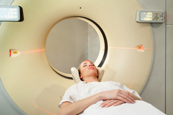
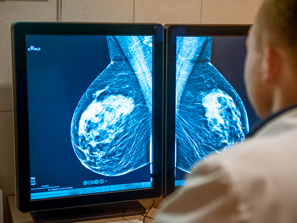
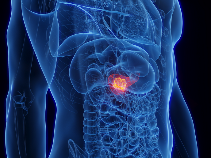
Imaging technology can be crucial for detecting at an early stage potentially fatal diseases such as cancer. In recent years, two-dimensional (2D) positron emission tomography (PET) scanning was replaced by more powerful 3D PET scanning, combined with computerised tomography (CT) scanning. However, scattered radiation resulting from increased sensitivity compromises accuracy, opening the door for improvements regarding this technology. The EU-funded project 'Accurate Reconstruction in PET: Fully 3D PET reconstruction with compressed scatter system matrix' (Acripet) worked on new technology to improve scanning capabilities. It developed a more accurate 3D system using a new system matrix to reconstruct the imaging process. This new method employs compression technology that reduces image 'noise' based on multi-node computer architecture, while remaining cost-effective and clinically feasible. The idea is that new computer power has the potential to improve PET image accuracy considerably, making it possible to diagnose cancer and similar diseases at an earlier stage. With this in mind, new particle tracking algorithms were developed and applied to the software needed. The process of photons entering detection systems was also improved, as were the speed of calculation and memory storage. All these improvements and upgrades led to better reconstruction of images during preliminary trials. Overall, project research and results have advanced the cause of highly accurate PET imaging greatly, which will be of great benefit in cancer diagnosis and patient follow-up. The technology looks very promising as well for patients within the fields of neurology and cardiology. The results may even be applied to small animal scanners, improving pre-clinical imaging used to develop pharmaceuticals and treatment.