Hi-precision breast cancer biopsy
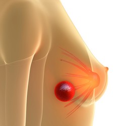
Breast cancer diagnosis is performed using clinical imaging modalities such as mammography and ultrasound. However, nearly 2 % of women with abnormal findings require a biopsy to confirm the results. Breast biopsy is an image-guided procedure that is time-consuming and of limited accuracy. Ideally a biopsy procedure should be comfortable for the patient with minimal risk of injury. To improve the overall performance of the technique, scientists on the EU-funded MAMMOCARE(opens in new window) (Breast biopsy system guided by positron emission mammography allowing real-time 3D visualization of tumour lesion and needle insertion guidance for higher sampling accuracy and efficiency) project developed a system based on positron emission mammography (PEM). This novel technology offers high sensitivity and spatial resolution. to detect small lesions that are difficult to find with conventional imaging technologies. To achieve its goals, the consortium integrated the expertise of three small- and medium-enterprises in PEM imaging technology. The MAMMOCARE system allows real-time three-dimensional visualisation of the lesion and guidance of the needle. The system allows the preoperative calculation of the optimal needle path. The high precision mechanics of the biopsy positioning module reduces inadequate sampling and positioning errors associated with other methods. During the initial part of the project, the teams worked closely to ensure that all operational, security and ergonomic aspects of the system design are integrated with the mechanical parts. Considerable efforts have also gone into the design of the software components for system control and interoperability. Following research and development activities, partners completed the clinical protocol and defined the patient inclusion and exclusion criteria for clinical validation. In this preliminary validation, eight patients scheduled for neo-adjuvant chemotherapy were scanned on the MAMMOCARE prototype system. Tumour visualisation on MAMMOCARE was appropriate for biopsy in five out of eight patients. In other cases, the uptake of the radioactive tracer (FDG) was too low for precise tumour visualisation (conventional PET/CT was also not able to visualise the breast tumours in these cases). The PEM system prototype for breast cancer imaging and biopsy was developed, validated, and is ready to enter the market. PEM-guided imaging is anticipated to reduce the duration of the biopsy procedure and benefit women through prompt and more accurate diagnosis.