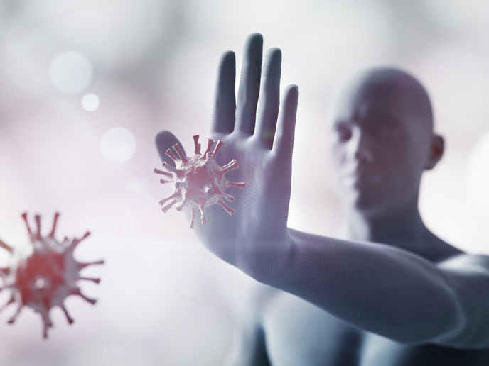
Cancer surgery reaches unprecedented levels of precision

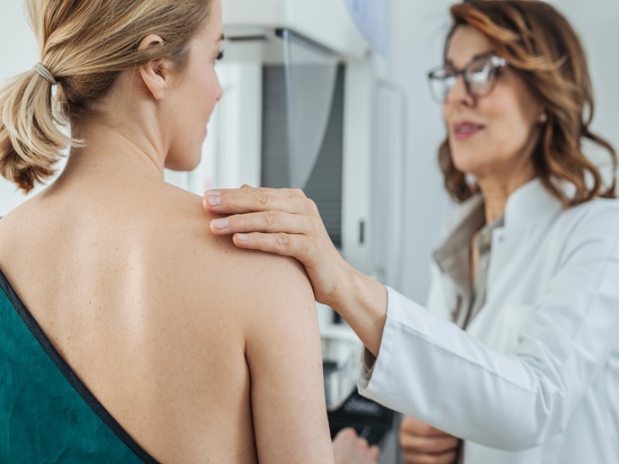
In oncological surgery, the challenge for the surgeon is to remove all the cancerous tissue while preserving as much as possible the healthy surrounding tissue. This crucial stage determines cancer recurrence, post-operative functionality and cosmetic outcome. Patient care is improved when immediate margin assessment is made available during surgery. The EU-funded HistologTM Scanner project, now known as Histolog® Scanner, offers a novel imaging modality to support clinicians in this decision-making process, improving patient quality of life and decreasing rates of reoperation.
Establishing a new approach in the operating room
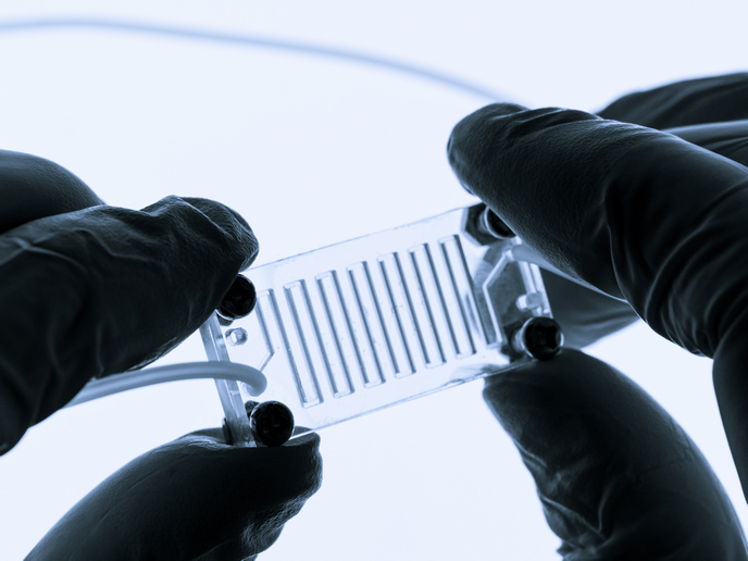
Tumour excision is the main treatment for a series of cancers, with the most commonly diagnosed one in Europe being breast cancer(opens in new window). In case this exceptionally delicate process results incomplete, the patient needs reoperation – a stress increasing and high-cost development. Based on massive parallel confocal microscopy, Histolog® Scanner(opens in new window), a CE mark product since 2016, offers an optimised intra-operative imaging technique that reduces this need. It enables real-time visualisation of cancerous cells on a surgical specimen during the operation process, while the morphology of the freshly excised tissue is immediately visualised without freezing or fixing. “Our immediate mission aims at setting a new standard for breast margin assessment, and to be applied in other cancer surgery applications, such as for the prostate or neurosurgery, because the underlying detection method applies to all cancerous pathology,” explains Bastien Rachet, CEO and co-founder of SamanTree Medical. “We are also developing an AI-based guidance software, which will provide an even stronger support to clinicians and further aid the standardisation of our approach.”
Medical innovation as an obstacle race
During the three-year HORIZON 2020 funding, the project team focused on generating clinical data required for the commercialisation of the patented platform. In the course of a clinical study performed in collaboration with Gustave Roussy Institute, one of the world’s leading cancer centres, we developed a set of Histolog® images of breast cancer specimens, in which a board of expert pathologists has annotated and classified both normal and cancerous tissue structures. This work was essential for the company to develop training material for clinicians. “The COVID-19 sanitary crisis would have compromised this project without our determination to keep it running,” admits Rachet. The team developed IT tools to support efficient remote work configuration with their international partners and proceed safely even amid the pandemic. “This example highlights the importance of proposing innovation in digital tools for the medical community and the crucial role of the financial support of such innovation by the European Commission,” emphasises Rachet.
Supporting cancer treatment in all its phases
So far, the innovative solution addresses the challenges faced in the operating room. The platform, though, has the potential to reinforce and complement pre-therapy diagnostics as well as lab analysis. Biopsy and specimen examination will also be considerably benefitted. “Enhancing the decision-making capabilities during surgery is only the beginning; we are also aiming to provide improved diagnostic capabilities to clinicians when performing biopsies or analysing specimens in the laboratory,” Rachet concludes. “We have already laid the groundwork for future collaborations in Europe and the United States, and we plan to move in this direction.”