

ESA scientists help cancer diagnosis with new X-ray camera
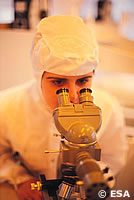
European Space Agency (ESA) scientists developing devices to capture X-rays from objects in space have designed a camera that could become a powerful new weapon in the fight against cancer. Members of the Science Payloads Technology division of the research and science support department, at ESA's science, technology and engineering research centre (ESTEC) in the Netherlands, have developed a small, lightweight X-ray camera that can aid cancer diagnosis and be used for continuous monitoring during operations. Effective treatment of cancer relies on the early detection and removal of cancerous cells. At this stage, the cancer is very hard to spot, and current medical equipment can give doctors only limited information on tissue health. In the case of breast cancer, cancer cells tend to congregate in the lymph nodes, from where they can rapidly spread throughout the rest of the body. A surgeon must perform an exploratory operation to try to identify and remove the diseased tissue. If identification is not possible, the doctor may be forced to remove the whole of the lymphatic system, which causes serious disruption to the body's hormonal balance. The new camera works by picking up images from a radioactive tracer injected into or near the breast tumour. The small size of the camera makes it possible to continuously image the cancerous tissue during surgery. 'There is no photography involved in the camera we envisage. It will be completely digital, so the surgeon will study the whole lymphatic system and the potentially cancerous parts on his monitor. He then decides which parts he removes,' says Dr Tone Peacock, Head of the Science Payloads Technology division. The discovery was made while the ESA team were developing a camera which could create images from high energy X-rays, as many celestial objects give out large quantities of X-rays but little visible light. ESA's XMM Newton X-ray telescope, currently in orbit, observes only low energy X-rays. For the first time, the ESTEC researchers have produced a microchip capable of detecting hard X-rays instead of visible light. Rather than silicon, the new chip is made from a chemical compound called epitaxial gallium arsenide. This material, developed under the ESA leadership of Dr Marcos Bavdaz, has successfully completed extensive tests at German X-ray test facility HASYLAB. Having made the basic camera sensor, the next stage will be to develop a system to send the images to television screens in real time. 'We are developing that now with our industrial partners, such as Metorex, a research and development company in Finland,' says Peacock. Once ESA has developed the technology to make the X-ray camera work, its task is done. The industrial partners will take over to produce a camera for medical use. ESA will adapt its design to provide European astronomers with a new view of the Universe.