Marching toward genetic resolution
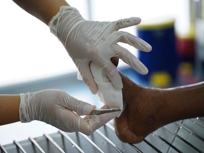
Microbial cells vary genetically, biochemically, physiologically, and in their behaviour in general. The key to understanding their properties lies in the unravelling of the finer detail of their structure and their interactions with each other. Major advances have already been made in this field, and now, even more impressive is the use of nano robots in cellular study. The development of nanoscale robots, just a few cm3 in size, will allow the retrieval of information on the fundamental cellular processes such as cell growth, cell proliferation and communication. The robots utilise AFM (Atomic Force Microscope) technology which allows the imaging of materials at the atomic level. AFMs use a ceramic or semiconductor tip one atom wide positioned at the end of a cantilevered bar. As the tip is moved over the material, it either continuously touches or periodically taps the surface and bends as it is repelled or attracted to the structure. A laser picks up the deflections. In this way topological data can be obtained with nanometric resolution. Additionally surface properties can be assessed with regard to elasticity, conductivity, friction and other physical properties. One key advantage of the use of AFM technology over other microscopes which require dry samples is that live samples can be used. This means that cells can be studied in their natural environments and that processes can be followed in real time. In such real time environments, the microrobot can operate in a scanning mode whereby an image of a solid surface is obtained. Alternatively, it can perform nanoindentation whereby liquid samples can be imaged through repeated measurements throughout the sample. Using a cluster of robots simultaneous measurements can be made in different parts of the bacteria. Such a detailed, complete imaging tool is set to provide the answers to many as yet unsolved genetic questions. All that remains is for the army of robots to set off.