Imaging spatial attention awareness in the brain
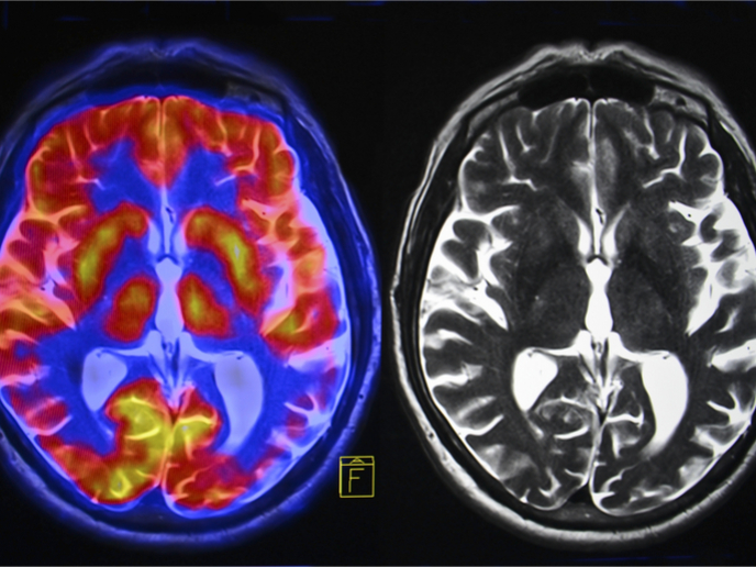
The 'Multimodal imaging of spatial attention networks in the human brain' (Multimodalattention) project chose three techniques to study spatial attention networks in the living brain. Specially selected technologies allow each set of information to complement the other sets. This presents a full picture of what is going on in the right hemisphere of the live brain. Diffusion tensor tractography (DTT) allows the study of white matter connections in the brain. Complementing this method is functional magnetic resonance imaging (fMRI), giving a picture of the dynamics over a shorter timescale. Finally, electroencephalography (EEG) gives sensitivity to interactions invisible to fMRI methods. The Multimodalattention team observed that the areas of the occipital lobe responsible for visual processing and the parietal lobe for determining spatial sense and navigation are dominant in the right side of the brain. They also found that there is visuo-spatial attention dominance in the right hemisphere due to quicker processing than in the left side of the brain. Effects of disorders in the right hemisphere are debilitating. An example is neglect where patients do not take into account information coming from the left side of space. Two other phenomena related to right hemisphere damage are extinction, and simultanagnosia where stimuli presented at the same time are sometimes not perceived. Patients with right-hemisphere damage suffer severe functional impairment in everyday activities. The Multimodalattention research results will help clinicians to predict recovery and plan patient-specific therapies.


